If you enjoy this content, please share it with a colleague
Philips
RELATED CONTENT
A new report from KLAS, “Digital X-Ray Performance 2013: Impact of a Wireless Workflow,” shows providers are seeing big benefits from wireless detector technology. With all of the major vendors offering it and prices coming down, wireless digital radiography (DR) is becoming a standard for digital X-ray room replacements.
Philips Healthcare announced that CX50 xMatrix, the first portable ultrasound with Philips' Live 3-D transesophageal echo (TEE), now offers 2-D intracardiac echo (ICE) capability. The CX50 xMatrix with available Live 3-D TEE and ICE was first shown in Paris at the EuroPCR in May.
If you are part of a health system that has spent months building and designing a new picture archive and communications system (PACS), it is undoubtedly an exciting time. Reaching the point of PACS activation and getting staff up and running is a true milestone. Once your company reaches this point, it may feel like the hard work is over and that it is time to take a deep breath, but in reality there is still much more to do and questions that have to be answered in order to fully support your organization during and post PACS go-live.
During the American College of Cardiology 2013 (ACC.13) annual meeting in March, vendors discussed several trends they are observing in the cardiac ultrasound market and displayed the latest echo advances.
Royal Philips Electronics showcased its latest addition to the ClearVue ultrasound family of products at the American College of Obstetricians and Gynecologists 61st Annual Clinical Meeting in New Orleans May 4-8, 2013. The ClearVue 650 is a lightweight and cost-effective system that spans a range of applications from obstetrics and gynecology to cardiology, abdominal, vascular, breast, musculoskeletal, urology and general imaging.
The Focused Ultrasound Foundation, Royal Philips Electronics and InSightec Ltd today announced that they are collaborating with leading clinicians in the field and support the establishment of a multicenter registry to evaluate MR-guided focused ultrasound treatment for uterine fibroids (also known as uterine leiomyomas).
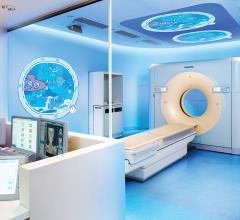
Imaging children has challenges that relate to the size, maturity and anxiety of the patient. In addition, we have become increasingly aware of the risks associated with radiation exposure, an issue that is of paramount importance to young patients. Through the efforts of professional organizations, industry and the Image Gently campaign, launched in 2008 by The Alliance for Radiation Safety in Pediatric Imaging, tremendous advancements have been made in technology and its use to help meet these challenges.
New computed tomography (CT) dose studies and growing public media attention have made minimizing unnecessary radiation dose to patients a priority for medical imaging facilities. In addition, state regulatory agencies and accrediting bodies are increasing their oversight and regulation of radiation dose. Reducing dose while maintaining good clinical image quality, however, is complex.
At the International Society for Magnetic Resonance in Medicine’s (ISMRM) 21st Annual Meeting in Salt Lake City, Utah, Philips Healthcare demonstrated a range of clinical functionalities in magnetic resonance imaging (MRI).
When every second counts in clinical situations such as those encountered in the emergency room and trauma centers, fast high-resolution CT scanning at low radiation dose helps radiologists render quick, confident diagnoses while keeping in mind the patient's needs. Delivering speed and performance, Philips iDose4 generates results in seconds rather than minutes, with a majority of reference protocols reconstructed in 60 seconds or less. Moreover, the Metal Artifact Reduction for large Orthopedic Implants (O-MAR) reconstruction technology reduces artifacts from metal objects, thus reducing the potential loss of critical anatomical information.
Philips has taken CT scanning to a whole new level by developing medical imaging technologies that combine fast reconstruction, high image quality and low radiation dose. This advancement was brought about by Philips collaborating with Intel to enhance its imaging software to take advantage of state-of-the-art, massively-parallel computing platforms based on Intel Xeon processors.
This paper details the high performance derived from Philips reconstruction technologies: Philips iDose4 and O-MAR. Some of the software optimization techniques used by Philips engineers are also described. As a next step in transforming care, Philips has launched the next innovation, called IMR. IMR is a model-based iterative reconstruction, which will run on even more powerful Intel processors and pave the way for further advances in CT imaging.


 June 11, 2013


