If you enjoy this content, please share it with a colleague
Philips
RELATED CONTENT
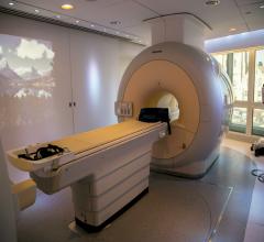
The average age of installed MRI scanners in the United States has increased from 8.7 years in 2010 to 11.4 years in 2013, according to a new market research report by IMV Medical Information Division.
Several industries have used cloud solutions for many years, but cloud computing only recently started to be used in healthcare. According to the National Institute of Standards and Technology (NIST), cloud computing is defined as “a model for enabling convenient, on-demand network access to a shared pool of configurable computing resources (e.g., networks, servers, storage, applications and services) that can be rapidly provisioned and released with minimal management effort or service provider interaction.”1 As more and more healthcare organizations (HCOs) adopt electronic medical records (EMRs), the cloud database has offered an efficient solution for image sharing, particularly in radiology where it is bridging the gap between referring physicians and radiologists.
The advent of hybrid PET/MR in 2011 brought the promise of vastly improved imaging technology in the form of a new modality that combined whole body positron emission tomography (PET) with magnetic resonance (MR) technology. Following two years of using PET/MR, we are seeing clinical benefits with this system at the University Hospitals Seidman Cancer Center in Cleveland.
Digital radiography (DR) has become a mainstay within many hospitals and radiology practices. The increased adoption of DR can be attributed to X-ray vendors dropping their prices, as well as the introduction of wireless DR, which offers more flexibility and improved workflow than fixed-plate DR. Now, many radiologists are opting to invest in DR systems rather than retrofit older computed radiography (CR) systems. While companies such as Samsung are just entering the DR market with new introductions, the focus today is not as much on introduction as it is on refinement. From smaller and lighter detectors for specific applications, to the development of features for dose monitoring and recording, DR is evolving to become more efficient for radiologists.
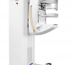
As breast density becomes a more recognized risk factor for breast cancer, Royal Philips announced 510(k) clearance from the U.S. Food and Drug Administration (FDA) for the Spectral Breast Density Measurement Application for its MicroDose SI full-field digital mammography (FFDM) system. The application is the first spectral Breast Density Measurement tool, meaning adipose (fat) and glandular tissue can be differentiated to accurately measure volumetric breast density.
Philips Healthcare introduced the IQon Spectral computed tomography (CT) system at RSNA 2013. It is the world’s first spectral-detector CT system built from the ground up for spectral imaging. It uses color to identify the composition of an image without involving time-consuming protocols.
Philips unveiled the Vereos PET/CT fully digital positron emission tomography/computed tomography (PET/CT) imaging system at the Radiological Society of North America Annual Meeting (RSNA 2013) in Chicago.
Royal Philips Electronics and Sectra announced an agreement to extend the term of their existing picture archiving and communication system (PACS) support partnership agreement.
Physicians have been utilizing conventional ultrasound, also known as b-mode ultrasound, for diagnostic imaging since the 1970s. However, over the past 10 years there have been significant technological improvements within the equipment, as well as development of new technologies that allowed ultrasound to become more widely adopted. Ultrasound equipment has gotten physically smaller, generates less heat and has become more power efficient. These upgrades, along with vast enhancements in image quality, have pushed ultrasound into the point-of-care setting. Point-of-care ultrasound has become widely performed in emergency rooms, PCP offices and obstetric practices. As healthcare reform continues to favor the use of more cost-effective solutions, this trend is expected to persist until ultrasound is used in every doctor’s office.
With concerns about radiation dose and reducing unnecessary imaging scans, advances in computed tomography (CT) systems have brought about technologies such as iterative reconstruction software, intraoperative capabilities and dose-tracking software. In addition, recent studies on the use of CT on select patient populations and the modality’s benefits in detecting certain cancers are showing that the risks of CT imaging can go both ways. While CT exams can add to a patient’s lifetime exposure to ionizing radiation, they can also be more beneficial in cases where magnetic resonance imaging (MRI) or ultrasound might not be able to detect early-stage cancers. Some of these trends in utilization indicate that appropriate low-dose CT imaging will be key across patient populations.


 February 06, 2014


