If you enjoy this content, please share it with a colleague
Philips
RELATED CONTENT
Wide bore magnetic resonance imaging (MRI) systems have allowed radiologists to offer patients the optimized comfort of conventional open bore systems, as well as the high-quality imaging of conventional closed bore systems. Because wide bore MRIs have broadened the demographic of patients who can be tested, the systems have gained widespread adoption in use, with many practices opting to equip their offices solely with wide bore systems.
Philips’ MobileDiagnost wDR system allows staff to bring radiography machines directly to patients, optimizing patient comfort. The detector technology and reduced need for retakes inherent to these machines contribute to the patients receiving a low X-ray dose, further enhancing patient experience.
There were three key trends in X-ray vascular imaging systems made evident from new products showcased at the Radiological Society of North America (RSNA) annual meeting in late 2012. These include new hardware and software to lower ionizing radiation doses and improve image quality, better dose tracking software, and enhanced maneuverability for improved patient access and use in the growing hybrid OR market. As interventional procedures become more complex, imaging times have increased, raising concerns that are now being addressed by vendors.
October 8, 2013 — Philips Healthcare kicked off Breast Cancer Awareness Month at its global healthcare headquarters in Andover, Mass. As part of its campaign, Philips is driving a mammogram truck on a three-month, 10,000-mile journey to visit hospitals and clinicians in more than 13 U.S. markets.
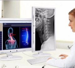
Philips released the Philips Clinical Review Display, a new solution for the fast-growing medical facilities market in Hong Kong. With aging population a worldwide phenomenon and computerized imaging systems for health diagnostics rapidly developing, the demand for quality clinical displays is increasing.
Royal Philips and Accenture announced the creation of a proof-of-concept (POC) demonstration that uses a Google Glass head-mounted display for researching ways to improve the effectiveness and efficiency of performing surgical procedures. The demonstration connects Google Glass to Philips IntelliVue Solutions and proves the concept of seamless transfer of patient vital signs into Google Glass, potentially providing physicians with hands-free access to critical clinical information.
Philips Speech Processing Solutions announced it will partner with ZyDoc, a medical transcription, speech recognition and natural language processing (NLP) technologies integrator, to continue to provide advances in voice productivity solutions.
The addition of magnetic resonance (MR) imaging and spectroscopy to positron emission tomography (PET) is more expensive and more technically challenging compared with PET/computed tomography (CT). PET/CT is successful because the inclusion of CT has major advantages: accurate lesion localization, the identification of non-PET avid lesions and effective attenuation correction in a rapid, efficient combined examination. The addition of CT is particularly valuable for lungs and liver, where fluorodeoxyglucose (FDG) PET is limited by spatial resolution and relatively low target-to-background differential biodistribution. Presumably, PET/MR may disclose unique important diagnostic and prognostic information in selected patient groups.
With concerns about radiation dose and reducing unnecessary imaging scans, advances in computed tomography (CT) systems have brought about technologies such as iterative reconstruction software, intraoperative capabilities and dose-tracking software. In addition, recent studies on the use of CT on select patient populations and the modality’s benefits in detecting certain cancers are showing that the risks of CT imaging can go both ways. While CT exams can add to a patient’s lifetime exposure to ionizing radiation, they can also be more beneficial in cases where magnetic resonance imaging (MRI) or ultrasound might not be able to detect early-stage cancers. Some of these trends in utilization indicate that appropriate low-dose CT imaging will be key across patient populations.
The use of 3-D echo can help improve the accuracy and reproducibility of cardiac quantification. The technology has the advantage of removing the inter-operator variability by imaging whole volume datasets of the heart, so specific images or organ views can be extracted and reconstructed in any position, similar to CT or MRI datasets. Also, because a volumetric dataset is captured, exam times can be shortened, instead of spending time trying to get just the right angle for a 2-D slice view. Cardiac quantification can also be improved by measuring the entire heart or ventricle, rather than just slices of it. New software also automates this quantification.


 November 04, 2013


