If you enjoy this content, please share it with a colleague
Philips
RELATED CONTENT
Elekta and Philips Healthcare announced The University of Texas MD Anderson Cancer Center (Houston, Texas) has signed an agreement to join a research group to advance the development of an innovative image-guided treatment technology for cancer care. The technology merges radiation therapy and magnetic resonance imaging (MRI) technology in a single system. MD Anderson is the second member of the research consortium, which will comprise leading radiation oncology centers and clinicians, and already includes the University Medical Center Utrecht (the Netherlands).
According to a new market research report "Breast Imaging Technologies Market (Digital Mammography,3D Breast Tomosynthesis, Breast MRI, Breast Ultrasound, Molecular Breast Imaging, Optical Imaging, PET/CT/PEM Modalities) Technology and Market Analysis & Global Forecasts to 2017" is an attempt to showcase the market impact of current and emerging breast imaging technologies having excellent growth potential in the coming five years. The technologies profiled in the report are segmented into Ionizing breast imaging modalities and Non-Ionizing breast imaging technologies on basis of radiation. Ionizing breast imaging modalities include Mammography, 3D Breast Tomosynthesis, Cone beam Computed Tomography (CBCT), Positron Emission Mammography (PEM), Molecular Breast Imaging (MBI), Positron Emission Tomography (PET) and Breast Specific Gamma Imaging (BSGI). The various Non-ionizing modalities for breast screening covered in the report are Breast MRI, Optical Imaging, Breast thermography and Breast Ultrasound.
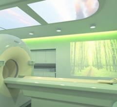
Royal Philips Electronics today announced a landmark agreement with the Farah Medical Complex in Jordan that will see the development of a new hospital which has patient experience firmly at the center of its ethos. Through this partnership, Philips will provide a customized package of 15 advanced imaging solution systems, healthcare informatics and services, in addition to a suite of 20 Ambient Experience rooms, the largest installation to date in any hospital worldwide.
iCAD, Inc. (NASDAQ: ICAD), an industry-leading provider of advanced image analysis, workflow solutions and radiation therapy for the early identification and treatment of cancer, today announced approval by the U.S. Food and Drug Administration (FDA) for use of the company’s next generation mammography computer-aided detection (CAD) platform, PowerLook Advanced Mammography Platform (AMP), with Digital CAD for Philips’ MicroDose Full-Field Digital Mammography System.
December 20, 2012 — At the 98th annual meeting of the Radiological Society of North America (RSNA), Royal Philips Electronics continued the Imaging 2.0 journey by showcasing several new features for existing image modalities that deliver clinical benefits to customers while simultaneously answering the economic challenges clinicians face worldwide.
The latest advances in cardiovascular imaging are usually shown first at the Radiological Society of North America (RSNA) annual meeting, the largest radiology show in the world, held the last week of November in Chicago. After spending five days walking three expo halls filled with more than 600 product vendors, the following is my editor’s choice for the most innovative new cardiovascular imaging technology.
There were several evident trends on the show floor at RSNA 2012, including interest in software fueled by Stage 1 and 2 meaningful use requirements, new mammography solutions and innovations in imaging hardware.
November 27, 2012 — At the 98th annual meeting of the Radiological Society of North America (RSNA) in Chicago, Royal Philips Electronics unveiled the company’s next chapter of its unique approach to radiology.
At ASTRO 2012, IBA featured a scale model install of its Proteus One compact proton system combined with the Philips ...
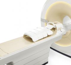
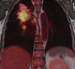
The last decade has seen a significant advancement in imaging technology due to developments in the hardware and software space. It was clear to the radiologists, clinicians and imaging scientists very early on that no single imaging modality, be it magnetic resonance imaging (MRI), computed tomography (CT) or positron emission tomography (PET) could meet all the needs of a clinician treating a patient.

 January 25, 2013


