If you enjoy this content, please share it with a colleague
Philips
RELATED CONTENT
June 22, 2012 — Philips introduced the iDose4 Premium Package, which includes two technologies that can improve image quality: the iDose4 iterative reconstruction technique, and metal artifact reduction for large orthopedic implants (O-MAR).
June 18, 2012 — United Health Services (UHS), a locally owned, not-for-profit, 916-bed hospital and healthcare system serving the greater Binghamton, N.Y. area has selected Philips Live 3-D transesophageal echo (TEE) technology for cardiac care, marking the system’s 2,500th installation in North America.
June 8, 2012 — At this year’s SNM Annual Meeting, June 9-13, Philips Healthcare is highlighting its portfolio of nuclear medicine (NM) applications for IntelliSpace Portal, adding to its existing portfolio of CT, MR and multimodality applications. The full suite of NM applications will now offer comprehensive processing and review capabilities at a level normally reserved exclusively for dedicated NM workstations. The company will also showcase solutions designed to increase diagnostic confidence, enhance patient comfort, augment physician confidence, simplify clinical workflow and reduce lifecycle costs.
Southern Ohio Medical Center (SOMC) is a 222-bed, rural, nonprofit hospital in Portsmouth, Ohio, that serves approximately 120,000 patients in the Appalachian area. The computed tomography (CT) department is a 24/7 operation. It has two Philips iCT scanners — one in the emergency department (ED) and the other in its medical imaging department — and it utilizes the iDose4 iterative reconstruction technique on iCT scans. Modifying imaging protocols for high image quality while achieving doses as low as reasonably achievable (ALARA) has become an endeavor of the imaging department at SOMC.
With the precision afforded by today’s radiation therapy delivery systems, treatment planning software that helps direct the process must keep pace. The treatment planning system provides a 3-D view of the tumor that facilitates decisions about treatment options and helps the clinical team develop the best possible plan. They are a means to achieving the end goal for the patient — perfectly targeted, appropriate dose.
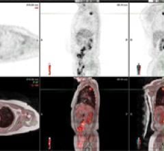
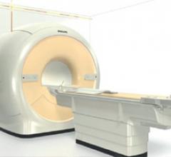
Magnetic resonance/positron emission tomography (MR/PET) is the newest imaging modality falling within the category of hybrid imaging. It sits at the crossroads of successful tradition and innovation. It is traditional in that it combines anatomical and functional molecular imaging from two potent imaging modalities; at the same time it is innovative, in that it combines the full potential of anatomic and multiparametric imaging of MRI with the molecular information of PET.
May 16, 2012 — Philips Healthcare has established a five-year agreement with St. Joseph’s Hospital and Medical Center in Phoenix to pursue research to help accelerate the advancement of magnetic resonance imaging (MRI) technology.
Single photon emission computed tomography (SPECT) remains a well-entrenched imaging modality for nuclear myocardial perfusion imaging (MPI) more than 30 years after its introduction. Due to SPECT’s reliability, cost-effectiveness and the wealth of data showing its clinical validation, it remains more common in MPI than its competition, positron emission tomography (PET).
Hardware and software advances are enabling echocardiography to greatly expand its capability with increased quantification accuracy, ease-of-use, increased workflow efficiencies and wider use outside of echo labs. Today, cardiovascular ultrasound systems are being integrated into point-of-care for triage, and in operating rooms and cath labs for procedural guidance to cut the use of contrast and ionizing radiation.
May 9, 2012 — Philips is bringing its newest computed tomography (CT) to St. Francis Hospital in Poughkeepsie, N.Y., home to the area’s only Level II trauma center.


 June 22, 2012


