If you enjoy this content, please share it with a colleague
Philips
RELATED CONTENT
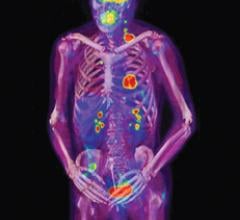
Since it was named “Invention of the Year” by TIME magazine in 2000, positron emission tomography/computed tomography (PET/CT) has been hailed as a winning combination. It captures anatomical information from the CT and functional information from the PET to create a fused image at once, reducing some of the challenges that occur in fusing two images acquired at separate times from separate modalities.
Musculoskeletal impairments are reported by more than one out of every four Americans, and in 2008 an estimated 46 million, or one in five adults, reported arthritis.1 Local injections of therapeutic agents into articular structures can lead to rapid decreases in pain and inflammation without many of the serious side effects associated with systemic medications. However, as with any diagnostic or therapeutic procedure, success depends on the expertise of the clinician and the accuracy with which the medications are injected into the affected joint space. Ultrasound is an emerging imaging modality which affords dynamic, real-time, cost-effective and physician-controlled visualization of anatomic impairments. Recent data has demonstrated that the use of ultrasound imaging improves accuracy rates in joint injections.
Multimodality imaging in medicine can provide a physician with tools for making an accurate diagnosis prior to making treatment recommendations and helps the physician to lessen the potential for restaging, simplifying the image evaluation process and improving patient care in the future. The benefits also can be applied to the improvement of imaging in clinical trials, where precision and standardization is a necessity. For many diseases, including cancer, heart disease and certain brain disorders, the current and typical imaging process requires the acquisition of both positron emission tomography (PET) and computed tomography (CT) scans.
May 1, 2012 - Philips announces the release of EasyDiagnost Eleva DRF 5.0, now available with a wireless portable detector and detector sharing functionality. The device is Philips’ latest advancement in digital radiography (DR) and fluoroscopy, helping to bring more versatility and increased workflow efficiency to busy hospitals.
April 16, 2012 — Philips Healthcare announced it is collaborating with Brainlab AG to create a comprehensive intra-operative magnetic resonance imaging (MRI) solution with the goal of streamlining neurosurgery procedures. Ingenia MR-OR is based on Philips’ digital broadband Ingenia MRI system, 1.5T and 3.0T, and is designed to be combined with Brainlab’s integrated operating room (OR) solutions.
Philips Healthcare recently installed an Allura Xper FD 20/20 biplane angiography X-ray system at Primary Children's Medical Center in Salt Lake City, with a National Basketball Association (NBA) theme to put children at ease prior to and during interventional radiology procedures.
April 11, 2012 - Philips Healthcare announced that more than 100 purchase orders have been made for Brilliance iCT scanner in Greater China. iCT is the world's fastest operating CT scanner and has been in high demand among China's medical institutions for its high-end and cutting-edge technologies and outstanding performance in clinical tests ever since its late 2008 release into the Chinese market. In only three years, the innovative computer tomography scanner at the forefront of medical technology has been largely instituted among the most reputable medical care facilities throughout China.
In arc therapy, a linear accelerator gantry moves in a continuous arc around the target while delivering radiation dose. Patients have been routinely treated with this technology since the 1980s, when it was put into use for stereotactic radiosurgery (SRS) of the brain. The advantage was that the low-dose region was spread out over a larger amount of healthy brain, reducing treatment toxicity.
While computed tomography (CT) has revolutionized medical imaging, it has also required much higher doses of ionizing radiation than was previously used in traditional X-ray imaging. In recent years, vendors have turned their attention to developing technology to drastically reduce dose.
With the advent of handheld ultrasound systems, the idea of a doctor in the emergency department (ED) with an ultrasound clipped to his keyboard is not too far-fetched. We are not there yet, but competition is growing in the market. While there are signs the market has begun to mature, there also are signs it could expand into new territory.


 May 03, 2012


