If you enjoy this content, please share it with a colleague
Philips
RELATED CONTENT
Philips Healthcare announced the commercial availability of its high-performance IntelliSpace Universal Data Manager at the 2016 Radiological Society of North America (RSNA) annual meeting, Nov. 27-Dec. 1 in Chicago.
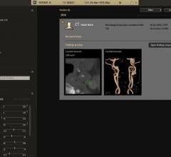
Philips announced the introduction of Illumeo, a new imaging and informatics technology with adaptive intelligence that enhances how radiologists work with medical images. The intelligent software is the first, according to the company, to combine contextual awareness capabilities with advanced data analytics to augment the work of the radiologist. Its built-in intelligence records the radiologists' preferences and adapts the user interface to assist the clinician by offering tool sets and measurements driven by the understanding of the clinical context.
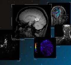
Philips announced the introduction of a suite of magnetic resonance (MR)-based software applications dedicated to neurology, which will debut at the Radiological Society of North America's (RSNA) 102nd Scientific Meeting and Annual Assembly in Chicago. With these introductions for its Ingenia family of digital MRI systems, Philips is reinforcing its global leadership in Neuro MR software applications, providing radiologists with the necessary tools to resolve complex health questions and explore new territories in neurology.
Philips introduced DoseWise Portal 2.2, a next-generation radiation dose management software platform for healthcare providers to record, track and analyze radiation exposure to patients and clinicians. The latest version of DoseWise Portal includes enhanced connectivity and informatics capabilities to address key challenges faced by radiology departments, such as managing dose exposure to ensure patient and staff wellbeing, and improving integrated access to patient information to deliver data-driven decision support.
The past decade has witnessed significant developments in ultrasound technologies, ranging from portable devices, wireless transducers to 3-D/4-D ultrasound imaging and artificial intelligence. Researchers and scientists are endeavoring on developing technologies that simplify diagnostic procedures, improve efficiency of clinicians and enhance image quality. These research and development activities focus on improving overall quality of patient care. In addition, manufacturers are placing an emphasis on implementing automation in premium-tier systems, portable devices and point-of-care (POC) solutions. The prime focus of vendors will be on offering cost-effective devices with growing innovation and competition in the global industry.
As hospitals begin replacing their first-generation 64-slice computed tomography (CT) scanners after a decade of use, there are several considerations evaluation teams should think about when looking at the newer-generation scanners. Chief among these is the idea that more slices makes for a better scanner — which CT experts say is not necessarily the case, and there are costs versus benefits to consider when looking at high-slice systems.
For all the benefits of medical imaging, most forms come with the inherent danger of radiation exposure. Public radiation exposure has increased significantly overall in the last 30 years, and according to a 2006 report from the National Council on Radiation Protection and Measurements (NCRP), medical imaging accounted for nearly half (48 percent) of all public exposure that year. Several high-profile cases of excessive radiation exposure due to medical imaging have brought the discussion into the public eye, which in turn has spurred greater focus on radiation safety among healthcare facilities and their various regulatory bodies.
IBA (Ion Beam Applications SA) and Philips announced in September the launch of a dedicated patient-centric Ambient Experience solution for the proton therapy gantry of IBA’s Proteus Plus proton therapy solution. The announcement makes the Philips solution now available for both Proteus One and Proteus Plus.
October 14, 2016 — Philips announced at The American College of Emergency Physicians' (ACEP) annual meeting that it has ...
October 13, 2016 — Philips announced that it will be displaying a variety of solutions aimed at increasing diagnostic ...

 November 29, 2016


