If you enjoy this content, please share it with a colleague
Philips
RELATED CONTENT
Treatment planning systems are at the heart of radiation therapy (RT) and the key to improved patient outcomes. Once image datasets are loaded and the tumors are identified, the systems develop a complex plan for each beam line route for how the therapy system will deliver radiation.
Philips announced it would be showcasing a variety of nuclear imaging solutions at the Society of Nuclear Medicine and Molecular Imaging (SNMMI) 2016 Annual Meeting, June 11-15 in San Diego.
Elekta and Philips announced the installation of a third high-field (1.5 Tesla) magnetic resonance-guided linear accelerator (MR-linac) system at University Medical Centre (UMC) Utrecht in the Netherlands.
As radiation therapy continues to evolve, new techniques and technologies are largely focused on maximizing the dose to the tumor site while protecting surrounding tissue as much as possible. Image guidance is a critical component of treatment planning for tumor delineation and gauging treatment response, and cone beam computed tomography (CBCT) has traditionally been the modality of choice.
ITN Associate Editor Jeff Zagoudis spoke with Richard Morin, Ph.D., Brooks-Hollern Professor of Medical Physics, Mayo Clinic, Jacksonville, Fla., and co-chair of the Image Wisely committee, about the latest efforts in computed tomography (CT) dose reduction:
The trend toward consolidation in the healthcare industry continues to climb, with U.S. hospital mergers and acquisitions at their highest since 1999.
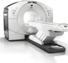
The last 12 months have seen significant growth in the positron emission tomography/computed tomography (PET/CT) segment of radiology, from both a manufacturing and a research perspective. One brand-new system received U.S. Food and Drug Administration (FDA) market clearance just months ago, while another started making its way into hospitals in the second half of last year. Several manufacturers also released software updates to help integrate PET/CT imaging into radiation therapy planning and execution. The increased interest was supported by several large studies exploring the advanced applications of PET/CT in oncology imaging.
Philips announced that any hospital or healthcare facility with one of its indicated computed tomography (CT) models can now become a lung cancer screening center.
Elekta, Philips and The Netherlands Cancer Institute, announced the installation of a 1.5 Tesla magnetic resonance (MR)-guided linear accelerator (MR-linac) system.
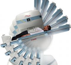
Introduced in the 1980s, magnetic resonance imaging (MRI) has become an integral part of any radiology department. The modality has most often been used for brain and spine scans, but new applications are expanding its capabilities in recent years. Vendors are also working to reduce both scan times and machine size for enhanced productivity.


 July 06, 2016


