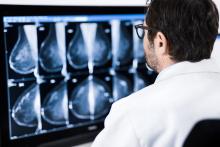
A combination of digital mammography and tomosynthesis detects 90 percent more breast cancers than digital mammography alone, according to a study appearing online in the journal Radiology.[1]

June 5, 2018 — The U.S. Food and Drug Administration (FDA) has cleared the Biograph Vision, a new positron emission tomography/computed tomography (PET-CT) system from Siemens Healthineers. The system offers new technologies to help improve image quality with motion management, improved delineation and quantification of small lesions, and new detector technology.

What something does is more important than what it is. This is common knowledge in consumer electronics


Breast imaging technologies have evolved rapidly in the last two decades to help physicians detect breast cancers at an earlier stage.


Value-based care and patient satisfaction are top priorities of radiologists across the field, from imaging technologists to radiation oncologists. For many practices, ensuring quality care requires a strategy involving many levels of patient interaction and care. What takes place behind the scenes — in the office, with clinicians and between different technologies — often makes the biggest difference.


The power to predict a cardiac arrest, support a clinical diagnosis or nudge a provider when it is time to issue medication — for many people artificial intelligence (AI) in healthcare represents a great new frontier. In addition to providing new applications improving delivery of care, AI is expected to bring improvements to hospital operations and to a range of clinical specialties.


A regional image exchange system is saving lives and reducing radiology costs in Maryland by improving the efficiency and effectiveness of diagnosing and treating stroke patients.


Sharon Gibbs, director of the radiology department at VCU Health in Richmond, Va., aims to provide quality, timely and efficient care. To do so, she must define, analyze and track the metrics and quality needs of her large care providing team, which consists of more than 40 faculty radiologists, over 200 technologists and numerous other stakeholders. 


The PACS — picture archiving and communication systems — have been in existence for more than 45 years. One of the first was created in 1972 by Richard J. Steckel, M.D., and the large-scale introduction occurred 10 years later at the University of Kansas. In the early ’90s, U.K. physician Harold Glass, M.D., pioneered the use of PACS to transform London’s Hammersmith Hospital into the first filmless hospital in the United Kingdom.


Subscribe Now