If you enjoy this content, please share it with a colleague
Philips
RELATED CONTENT
IBA (Ion Beam Applications S.A.) and Royal Philips announced the signing of an exclusive agreement to enhance access to proton therapy in India. The alliance combines IBA’s strengths in proton therapy and Philips’ expertise in clinical informatics and innovative imaging techniques for therapy planning and guidance.
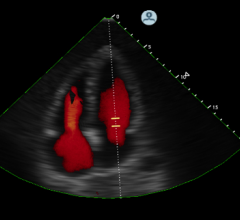
Royal Philips announced the introduction of HeartModelA.I., a new Anatomically Intelligent Ultrasound (AIUS) tool that brings advanced quantification, automated 3-D views and robust reproducibility to cardiac ultrasound imaging. Philips' fastest 3-D AIUS ultrasound measurement method was unveiled on its Epiq 7 ultrasound system during the American Society of Echocardiography (ASE) annual meeting in Boston.
More than 50 companies and organizations will display their latest products and services at the American Society of Echocardiography’s (ASE) 26th Annual Scientific Sessions, June 12-16 at in Boston, Mass. ASE 2015 is the world’s premier meeting for cardiovascular ultrasound practitioners, and promises a wealth of cutting-edge education, research, and the latest vendor technology.
In May, Group of Companies European Medical Center (GEMC) became the first private clinic in Russia to open a positron emission tomography (PET-CT) department producing its own pharmaceuticals. GEMC also opened a radiation cancer therapy department featuring two linear accelerators.
Today, treatment planning — the heart of radiation therapy systems — is almost entirely computer based, and is designed to help increase productivity by simplifying data for clinicians, making workflow smooth and seamless. This is key to helping improve patient outcomes. As technology progresses, so do treatment planning systems.
As one might expect, pediatric imaging presents a unique set of challenges compared to working with adult patients. Patient size, growth and development, and a distinct set of injuries and diseases all enter into the equation for pediatric radiologists.
A battle is taking shape in computed tomography (CT), one that pits the tubes that deliver X-rays against the detectors that record them. It is a fight over how to use X-rays of different energies — variations on the decades-old concept of selective energy CT (SECT), which promise to make diagnoses more accurate and patient management more effective.
As the saying goes, sometimes less is more — a maxim that is proving true in the world of medical imaging as remote viewing systems continue to advance. While some manufacturers are still utilizing software-based systems for reading and sharing imaging data, many are embracing browser-based models, otherwise known as zero-footprint viewers.
There have been several technology advances in PET/CT (positron emission tomography/computed tomography) systems recently introduced by vendors. On the show floors of the 2014 Society of Nuclear Medicine and Molecular Imaging (SNMMI) meeting and the 2014 Radiological Society of North America (RSNA) meeting, Philips, Toshiba and GE Healthcare introduced new PET/CT systems.
Proton Partners International Limited announced the appointment of global partners in its plans to build three proton beam therapy centers in the U.K. Partner organizations will provide state-of-the-art clinical equipment and technology solutions to the company.


 June 22, 2015


