News
Richard Wolf Medical Instruments Corp. and Maquet launched a non-exclusive sales partnership in the United States to offer OR integration solutions in the endoscopy market.
 February 03, 2014
 Blog
My generation has seen the rise of a frightening kind of STD, one that was unflinchingly lethal early on and since has been unrelenting, if survivable. Today AIDS is managed, not cured, the lethality of its cause, HIV, held at bay with a cocktail of daily medications. But now there is hope for a cure: radioimmunotherapy (RIT) promises the destruction of HIV-infected cells and the means to verify it.
February 03, 2014 
News
GE Healthcare, a unit of General Electric Company, developed features on the Logiq S8 general imaging ultrasound system. The innovations allow for enhanced image quality and streamlined workflow for healthcare providers, and help shorten exam times for patients. GE presented the advancements at the Radiological Society of North America (RSNA) 2013 conference.
February 03, 2014 Sponsored Content
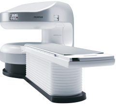
Fujifilm’s APERTO Lucent is a 0.4T mid-field, open MRI system addressing today’s capability and image quality needs ...
September 25, 2024 News
The Center for Tomography Research Laboratory (CTECH Labs) introduced its latest technology in brain scanning at the 6th International IEEE Engineering in Medicine and Biology Society Conference on Neural Engineering, San Diego, Calif., Nov. 7, 2013.
February 03, 2014 News
Medical Insight’s EasyViz was selected in a robust program initiated in Denmark where improved efficiency in patient care is being demonstrated. Two regions in Denmark have realized their strategic vision using a vendor neutral archive (VNA) paired with EasyViz, an enterprise diagnostic viewer for radiologists, clinicians, radiographers and more. Radiologists and clinicians in 14 hospitals can access clinical exams in seconds directly or from any Internet application. This eliminates the need and time for radiologists and clinicians to take geography, IT infrastructure and workstations into consideration.
February 03, 2014 Sponsored Content

News | Computed Tomography (CT) | August 06, 2024
SPONSORED CONTENT — Fujifilm’s latest CT technology brings exceptional image quality to a compact and user- and patient ...

Feature
Initial acceptance test results of ProTom International Inc.'s Radiance 330 Proton Therapy System are exceeding expectations of medical physicists charged with clinical commissioning at the McLaren Proton Therapy Center (MPTC) in Flint, Michigan.
February 03, 2014 Sponsored Content
News | Computed Tomography (CT)
SPONSORED CONTENT — Fujifilm’s latest CT technology brings exceptional image quality to a compact and user- and patient ...
August 06, 2024 
Videos | PACS
Carestream's Vue family includes RIS/PACS/archiving as well as vendor-neutral archives, physician and patient portals ...
February 03, 2014 News
Blackford Analysis, a provider of software products that accelerate comparison of medical images, introduced its products to the Middle Eastern market at the Arab Health Congress 2014 in Dubai. Designed to be integrated directly into any image viewer, such as a PACS, Advanced Visualization Viewer or Universal Viewer, Blackford Analysis’ products work within existing systems to enable instant comparison of multiple image studies.
February 03, 2014 
Feature | Greg Freiherr
Looking for signs of cancer in the mammogram of a dense breast is like looking for a polar bear in a snowstorm. Because they absorb X-rays, glandular and fibrous tissue appear white, as do microcalcifications, lumps and other lesions that may indicate breast cancer.
February 03, 2014 Sponsored Content
News | Artificial Intelligence
SPONSORED CONTENT — EnsightTM 2.0 is the newest version of Enlitic’s data standardization software framework. Ensight is ...
June 21, 2024 
Feature | Raissa Rocha
The practice of breast imaging and screening in women at risk for breast cancer is evolving as new imaging modalities are incorporated and researchers continue to study the implications of dense breast tissue in patients. At the 2013 annual meeting of the Radiological Society of North America (RSNA) in Chicago, there were several sessions highlighting trials and studies that brought breast density and cancer detection to the forefront.
February 03, 2014 Feature | Williette Nyanue
Several industries have used cloud solutions for many years, but cloud computing only recently started to be used in healthcare. According to the National Institute of Standards and Technology (NIST), cloud computing is defined as “a model for enabling convenient, on-demand network access to a shared pool of configurable computing resources (e.g., networks, servers, storage, applications and services) that can be rapidly provisioned and released with minimal management effort or service provider interaction.”1 As more and more healthcare organizations (HCOs) adopt electronic medical records (EMRs), the cloud database has offered an efficient solution for image sharing, particularly in radiology where it is bridging the gap between referring physicians and radiologists.
February 03, 2014 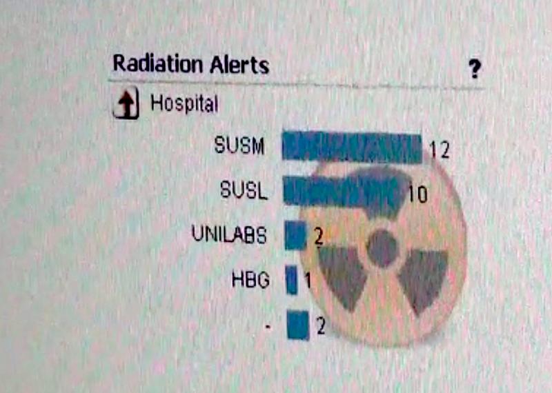
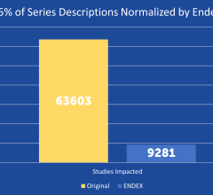
Feature | Mats Björnemo
Radiation dose continues to rise as the number of computed tomography (CT), nuclear, angiography and fluoroscopy examinations grow, leading to a greater risk of patient overexposure to radiation. Healthcare providers must reinforce their efforts to monitor and visualize dose levels from radiology examinations to enhance patient safety and meet new regulatory demands. There also is a need to justify and optimize the usage of radiation dose to find a balance between safer practice, image quality and lower dose — all for the benefit of the patient. Implementing tools for automatic and continuous follow up of radiation dose is at the forefront of meeting these challenges.
February 03, 2014 Sponsored Content

Feature | Artificial Intelligence
Did you know that approximately one-third of all the data in world is created by the healthcare industry and that ...
June 03, 2024 
Feature | Radiation Oncology | Williette Nyanue
The Elsie and Robert Pierson Radiation Oncology Center is a part of City of Hope National Medical Center — a dedicated, National Cancer Institute (NCI)-designated comprehensive cancer center located in Duarte, Calif.
February 03, 2014 
Feature | Pablo R. Ros, M.D., MPH, Ph.D.
The advent of hybrid PET/MR in 2011 brought the promise of vastly improved imaging technology in the form of a new modality that combined whole body positron emission tomography (PET) with magnetic resonance (MR) technology. Following two years of using PET/MR, we are seeing clinical benefits with this system at the University Hospitals Seidman Cancer Center in Cleveland.
February 03, 2014 Sponsored Content
News | Artificial Intelligence | June 21, 2024
SPONSORED CONTENT — EnsightTM 2.0 is the newest version of Enlitic’s data standardization software framework. Ensight is ...

Feature | Greg Freiherr
What was once diagnostic imaging is on track to becoming much more. Radiology is on the verge of a new era in which its focus goes beyond the traditional gatekeeper role, directing the earliest steps in patient management to one that adjusts the direction of care throughout the management of a patient.
February 03, 2014 
Feature | Williette Nyanue
Physicians have used radiation in medicine for more than a century. The use of radiation in diagnostic imaging, including computed tomography (CT), fluoroscopy, angiography, mammography, computed radiography (CR) and digital radiography (DR), as well as in nuclear medicine, has aided greatly in the diagnosis and treatment of cancer and other diseases.
February 03, 2014 Feature | Williette Nyanue
Digital radiography (DR) has become a mainstay within many hospitals and radiology practices. The increased adoption of DR can be attributed to X-ray vendors dropping their prices, as well as the introduction of wireless DR, which offers more flexibility and improved workflow than fixed-plate DR. Now, many radiologists are opting to invest in DR systems rather than retrofit older computed radiography (CR) systems. While companies such as Samsung are just entering the DR market with new introductions, the focus today is not as much on introduction as it is on refinement. From smaller and lighter detectors for specific applications, to the development of features for dose monitoring and recording, DR is evolving to become more efficient for radiologists.
February 03, 2014 
Case Study | Sponsored by Hologic
St. Peter’s Breast Center, located in Albany, N.Y, added 3D mammography (breast tomosynthesis) in 2011 because the staff knew it would improve diagnostic accuracy; even so, the technology has exceeded all expectations. In 2012 more than 22,000 mammograms were performed at the breast center.
February 03, 2014 Feature | Melinda Taschetta-Millane
To help identify trends and find out what topics Imaging Technology News (ITN) readers are interested in, our editorial team makes note of what the audience is viewing online at itnonline.com. In 2013, the topic of women’s breast health — specifically, breast density — dominated half of this list. Perhaps 2014 will be the year that standardized breast density reporting will become law in all states; 2013 got this off to a good start, helping to spread awareness. Other hot topics included healthcare reform and the role it could play in the radiology field, and of course breaking new technology.
February 03, 2014 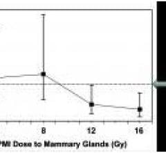
News
Survivors of breast cancer have a one in six chance of developing breast cancer in the other breast. But a study conducted in mice suggests that survivors can dramatically reduce that risk through treatment with moderate doses of radiation to the unaffected breast at the same time that they receive radiation therapy to their affected breast.
January 31, 2014 © Copyright Wainscot Media. All Rights Reserved.
Subscribe Now