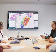
The Elsie and Robert Pierson Radiation Oncology Center is a part of City of Hope National Medical Center.
The Elsie and Robert Pierson Radiation Oncology Center is a part of City of Hope National Medical Center, a dedicated, National Cancer Institute (NCI)-designated comprehensive cancer center located in Duarte, Calif.
The center houses City of Hope’s department of radiation oncology, which includes the division of medical physics. The center is one of the largest radiation oncology centers in Los Angeles, seeing approximately 120-130 patients each day for treatment, imaging, consultation and follow-up.
The center is the result of an expansion that started in 2007 and was completed in the fall of 2012. The expansion doubled its space from approximately 14,000 square feet to almost 30,000 square feet, and allowed the department to integrate two cancer care trends together in one facility.
Combination of Imaging and Therapy
According to Jeffrey Wong, M.D., FASTRO, professor and chair, department of radiation oncology, City of Hope, the center was designed to bring technological advances in image guided radiation therapy (IGRT) and advanced imaging technologies in the areas of computed tomography (CT), magnetic resonance imaging (MRI) and positron emission tomography (PET) together in one location. “Technology advances in image guided radiation therapy allow for precise targeting of radiation energy, sculpting and conforming the dose of radiation to the target to minimize radiation effects to surrounding normal tissues and improve outcomes,” he said. By pairing IGRT and advanced imaging, the center was able to integrate the technologies and have them working together side by side.
This integration has allowed the center to apply imaging advances to the delivery of radiation therapy in a much more defined and immediate way. “The traditional workflow for radiation therapy has been primarily to use CT imaging to determine how best to target the radiation therapy,” Wong said. “Now we can incorporate into that workflow the use of PET imaging as well as MRI imaging, including multi-parametric MR imaging, to see things that sometimes the CT scan cannot visualize, such as better tumor definition and specific physiologic and chemical signatures that the tumor may emit.” Wong said that being able to detect tumors more accurately allows physicians to deliver radiation therapy more precisely, ensuring that they encompass the entire tumor, which improves outcomes and decreases side effects.
Tim Schultheiss, Ph.D., FACR, FAAPM, FASTRO, director, radiation physics, City of Hope, said the center has the GE Optima 560 16-slice PET/CT, which it utilizes for 4-D imaging as well as gating. The center also has a 3.0T MR, which has a number of modules for multi-parametric imaging. In addition, the center’s CT simulator is in the process of being replaced with a new 16-slice, large-bore CT scanner.
“When the CT simulator was first deployed in the field of radiation oncology, we really had no aspirations to produce diagnostic quality CT scans for purposes of treatment planning. We simply wanted to capture the patient electronically as a 3-D array of electron densities,” Schultheiss said. “But with the introduction of PET/CT and MR into the treatment planning process, we cannot and should not achieve images below the strictest diagnostic standards.”
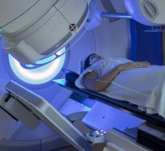
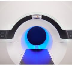
For treatment, the center utilizes two tomotherapy units from Accuray — which include one HD version — as well as Varian’s TrueBeam and TrueBeam STX. The TrueBeam systems are both equipped with an onboard imaging package that includes portal imaging and cone-beam CT, and are capable of gating. The center also has a high-dose-rate brachytherapy unit, a linear accelerator for total body irradiation and an intra-operative system that provides radiation therapy in a single fraction at the time of surgery for breast cancer patients.
Integration at Work
The integration of IGRT and advanced imaging is clearly seen in City of Hope’s 37-year-old bone marrow transplant program. The program is one of the largest in the world and transplants more than 500 patients per year. Physicians are currently researching the use of the TomoTherapy Hi-Art IGRT device, which sculpts the dose to the entire body but only targets the bone and bone marrow, in conjunction with CT and PET scans. Being able to target radiation to the bone marrow more precisely reduces doses to the normal organs when compared to whole-body radiation treatment.
According to Wong, the results of this research have been very encouraging. Physicians at the center have been able to offer this life-saving procedure to older patients and those who have advanced leukemia. “Patients older than 55 are now able to go through these types of procedures. The oldest patient to date treated with this type of technology is 70 years old,” he explained. “Also encouraging are results in patients who have no other bone marrow transplant options because their disease is too advanced.”
Wong said that with these new approaches physicians have been able to put these patients in remission. Some of the patients even continue to be in remission years later.
MRI-guided Focused Ultrasound
One technology that also is being evaluated at City of Hope is MR-guided focused ultrasound (MRgFUS). The center is one of only a few centers in the United States evaluating the technology in patients with cancer. Although the U.S. Food and Drug Administration (FDA) has approved MRgFUS for pain reduction in bone metastases — a technique already employed at the center — researchers are finding several additional areas in cancer treatment where the technology could be beneficial.
One such area is prostate cancer treatment. Using the ExAblate 2100 device made by InSightec Inc., researchers have found that MRgFUS has the ability to treat prostate cancer without the use of surgery or radiation therapy. “Instead of using radiation therapy to kill cancers, the device uses sound waves or ultrasound energy, and it focuses the energy to create a very tight focal spot of heat to ablate the tumor,” Wong said. He noted that what makes this technology exceptionally unique is that instead of using ultrasound images, which can sometimes be fuzzy or unclear, it uses MR imaging to target the tumor as well as for thermography. “Better imaging means more precise delivery of the treatment and the ability to potentially decrease risks and improve outcomes,” he said.
Scientists at City of Hope are also looking to MRgFUS to help translate discoveries in the laboratory to the clinic. “We have many scientists, including some associated with radiation oncology, who are developing targeted molecules that can carry therapy to tumors,” Wong said. “One of the challenges of this type of approach is you just can’t get enough of those agents into the cancer.” A possible solution may be found in the use of MRgFUS, which has shown promise in being able to increase the tumor uptake of those targeting agents. “Combining this technology with the biology from the lab can potentially make these agents more effective and help to translate those advances into the clinic,” Wong said.
Increasing Efficiency, Patient Volumes, Revenues
Wong said that the integration of IGRT and advanced imaging has also minimized the imaging that is used to take care of cancer patients and the exposure related to imaging tests, limiting retesting and associated costs. “When a person had images done in another part of the hospital or at other hospitals, we would often have to repeat those tests with the radiation therapy in mind, and patients would go through multiple tests just to get to the therapy state,” he explained. Now, with imaging and therapy in the same center, physicians can use the same imaging procedure for diagnostic purposes, therapy planning and to help target radiation.
Wong also noted the effects that integration has had on increasing efficiency and outcomes with bone marrow transplant patients. “Bone marrow transplant is an intensive procedure, and it can result in long hospital stays,” he explained. “But with a procedure such as targeted marrow irradiation with the tomotherapy unit, it’s better tolerated, and we feel it offers the potential to reduce side effects and shorten the hospital stay and the recovery time for these patients.”
Altogether, the integration has helped the center increase efficiency, patient volumes and revenues. “With the technologies, as we’ve acquired them, we’ve seen about a twofold increase in patient volume,” Wong said. “Along with that, we’ve seen a commensurate increase in revenue.”
Room for Growth
With these developments, Wong said there are many possibilities yet to be realized at the center. “We feel there is a potential for further increase in growth because we are one of the few centers where all these imaging technologies are in the same department of radiation oncology. I believe we are only one of perhaps a few radiation oncology departments in the U.S. that have MR-guided focused ultrasound. This technology platform is somewhat differentiating and will lead to growth,” he concluded.


 August 09, 2024

