Digital radiography (DR) has become a mainstay within many hospitals and radiology practices. The increased adoption of DR can be attributed to X-ray vendors dropping their prices, as well as the introduction of wireless DR, which offers more flexibility and improved workflow than fixed-plate DR. Now, many radiologists are opting to invest in DR systems rather than retrofit older computed radiography (CR) systems. While companies such as Samsung are just entering the DR market with new introductions, the focus today is not as much on introduction as it is on refinement. From smaller and lighter detectors for specific applications, to the development of features for dose monitoring and recording, DR is evolving to become more efficient for radiologists.
Wireless Detectors
Wireless detectors entered the U.S. market a few years ago, giving radiologists the versatility and mobility of CR detectors and analog film cassettes.
A report released in June 2013 from KLAS titled “Digital X-Ray Performance 2013: Impact of a Wireless Workflow,” showed that providers are continuing to reap big benefits from wireless detector technology. According to the report, providers saw increased efficiency in patient throughput due in part to the reliability and excellent image quality of wireless solutions. Providers are utilizing the ease and flexibility of wireless detectors, moving them between different rooms and X-ray systems. “It’s great to see wireless technology having a positive impact in healthcare,” said Brady Heiner, author and director of research at KLAS.1 “Seeing providers and vendors leveraging the wireless technology to create efficiencies that directly affect patient flow is win-win for all.”
With the success of wireless DR detectors, it is no surprise that vendors continue to enter into the market. Samsung made its debut into the DR market last year with the U.S. Food and Drug Administration (FDA) approval of its XGEO GC80 — complete with wireless options.
System Enhancements
Vendors are also adding features to existing DR systems and developing software to increase efficiency and workflow. Carestream introduced its bone suppression software, currently 510(k)-pending in the United States. It is designed to create a companion image to suppress the appearance of posterior ribs and clavicles while enhancing the visualization of soft tissue in the chest, making DR images easier to read.
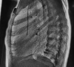
Agfa Healthcare announced the North American launch of its Musica2 catheter processing software last August. The software is designed to increase the visibility of peripherally inserted central catheter (PICC) lines and other low-contrast, tube-like structures such as endotracheal or feeding tubes in general radiology applications.
Smaller Detectors
DR detectors are also being tailored for specific applications. One trend featured at the 2013 Radiological Society of North America annual meeting (RSNA 2013) was the introduction of smaller and lighter format detectors for easier imaging of smaller anatomies. Carestream received FDA clearance this past June for its DRX 2530C wireless detector. The 25 x 30 cm detector is suited for pediatrics and the NICU, fitting into a tray under incubators so that radiology technicians can more easily obtain images. Similarly, Fujifilm Medical Systems USA Inc. also received 510(k) clearance for its gadolinium and cesium detectors for pediatric care in late 2013.
Low Dose
Another trend that is affecting every area of medical imaging is the development of products that aid in decreasing and managing radiation dose exposure. With DR, over-exposure often produces images that are a lot less noisy than under-exposed images. According to the RSNA 2013 presentation, “CR and DR Dose Reduction and Clinical Management,” by Charles E Willis, Ph.D., this has led to a gradual increase in exposure over time, and today radiologists often prefer, and opt for, images with higher exposure, as they are easier to read.2 With legislation regarding dose reporting and management already passed in two states and more to come, DR vendors are finding ways to decrease radiation exposure and maintain image quality.
Agfa HealthCare’s newest detector, the DX-D 35c, is an 11 x 14-inch wireless panel that uses a cesium iodide (CsI) scintillator with twice the detective quantum efficiency (DQE) of gadolinium-based detector technologies, delivering the potential for lower dose. Carestream’s bone suppression software also features IHE dose reporting, which collects dose information from all of the company’s DR and CR systems and distributes it to the healthcare providers’ picture archive and communication systems (PACS) for easy reporting. According to Titus, the dose-reporting feature is intended to streamline the collection of dose information from DR systems and support the ability to track dose values for each patient.
Point-of-care Use
As DR technology continues to improve, the use of DR systems in the point-of-care setting may be on the rise. This can already be seen with Konica Minolta’s launch of the ImagePilot Aero, an all-in-one digital imaging system for urgent care centers and community hospitals. The system, which combines the ease of use of the ImagePilot system with the high image quality of the Aero DR panel, offers private practices, imaging centers and community hospitals access to the productivity and versatility of wireless DR. As DR solutions get even smaller, lighter and easier to use, physicians can expect to see the use of DR in point-of-care continue to grow.
References
1. Fornell D, “Improving Efficiency and Throughput Using Wireless DR.” Imaging Technology News. 2013: 07:14-15.
2. Willis C, “CR and DR Dose Reduction and Clinical Management,”
session presented at RSNA 2013.


 November 26, 2025

