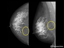
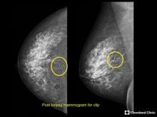
Mammography has been the gold standard for breast cancer screening ever since it was proven to reduce mortality in clinical trials in the 1960s. A 2015 report from the U.S. Department of Health and Human Services (HHS) revealed, however, that only 66.8 percent of women age 40 and over — the demographic traditionally targeted for screening — had received a mammogram in the previous two years.1 Understanding of fibroglandular breast density and its impact on cancer detection, as well as new screening guidelines that challenge conventional wisdom, are among the factors impacting compliance rates, and they suggest that screening has evolved beyond the one-size-fits-all approach of using mammography alone.


Recently, I was invited to participate in a roundtable discussion focused on uncovering the challenges and inefficiencies associated with stereotactic breast biopsies. In preparation, my colleagues and I spent a good deal of time reviewing patient case studies to identify a few that would best illustrate the issues related to this diagnostic procedure.


Healthcare spending is on the rise. According to a 2014 forecast by FMI, a market research group, total global healthcare spending will reach $52 billion by 2018 — less than two years away. Further research shows that nearly half of the money (40 percent) will be spent on equipment, an encompassing term that includes everything from beds to gurneys to imaging equipment. Radiology actually makes up the lion’s share of equipment costs, accounting for approximately 1/5 (depending on the type of facility) of total equipment expenditures, per a 2015 report from market research firm Medical Architecture Planning Systems. At the same time, it is also a major revenue driver for hospitals, with radiology expected to account for $49 billion of total global medical imaging revenue by 2020, according to BCC Research, a market research company.


As healthcare continues to march forward with numerous reforms, it falls to its various accrediting bodies to set standards to guide actual progress. Radiology is subject to many such accrediting bodies, but perhaps the largest is the Joint Commission, which oversees accreditation for all healthcare specialties. The most recent updates — including changes to the survey process itself as well as the scoring system — could have a major impact on radiology departments as a whole. Judith M. Atkins, RN, MSN, president and CEO of McKenna Consulting, spoke to radiology administrators at the 2016 annual meeting of the Association for Medical Imaging Management (AHRA) to help them parse through some of the latest updates and decide how best to address them.


Science fiction offers a lot of ideas for creating new body parts on demand, and the advancement of 3-D printing (also called additive manufacturing) is slowly translating this idea into science fact. Today, the 3-D printed anatomic models created from patient computed tomography (CT), magnetic resonance imaging (MRI) or 3-D ultrasound imaging datasets are used for education and to plan and navigate difficult procedures. These models are used to teach about complex or rare cardiac or congenital conditions that up until recently could only be seen using examples extracted from cadavers. Anatomical models of rare cardiac anatomy can be printed on-demand from CT scans of surviving patients.


I doubt any of us will forget 2016. Events, both tragic and bizarre, have owned the headlines and have provided more than a little water cooler fodder. As we close another year of activity within our own world of medical imaging, we too have had plenty to talk about. From acquisitions to rebranding; from new product releases to updated vendor platforms. There was nary a week that went by without something interesting hitting my feeds. Here are some important developments that I believe are making waves and setting the course for imaging, specifically enterprise imaging, as we move further into the second half of this second decade of the 21st century.


With the New Year nearly upon us, the diagnostic imaging industry will finally begin to realize the effects of Section 502 of the Consolidated Appropriations Act of 2016. For more than a year, the industry has been humming with talk of anticipated reduction of Medicare payments, expected to impact diagnostic imaging facilities still using analog and computed radiography (CR) technologies. As we are well aware, Section 502 of the Consolidated Appropriations Act of 2016 provides that Medicare reimbursements will be reduced by 20 percent per year for providers submitting claims for analog X-ray studies beginning this year. From 2018 through 2022, payments for imaging tests performed on CR equipment will be reduced by 7 percent, with a 10 percent reduction beginning in 2023.


Beginning Jan. 1, 2017, the Centers for Medicare and Medicaid Services (CMS) will begin enforcement of the Consolidated Appropriations Act of 2016, a piece of legislation that incentivizes healthcare providers to transition imaging departments to digital radiography (DR). 


The past decade has witnessed significant developments in ultrasound technologies, ranging from portable devices, wireless transducers to 3-D/4-D ultrasound imaging and artificial intelligence. Researchers and scientists are endeavoring on developing technologies that simplify diagnostic procedures, improve efficiency of clinicians and enhance image quality. These research and development activities focus on improving overall quality of patient care. In addition, manufacturers are placing an emphasis on implementing automation in premium-tier systems, portable devices and point-of-care (POC) solutions. The prime focus of vendors will be on offering cost-effective devices with growing innovation and competition in the global industry. 


Subscribe Now