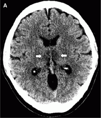
A, Image from noncontrast head CT demonstrates symmetric hypoattenuation within the bilateral medial thalami (arrows). B, Axial CT venogram demonstrates patency of the cerebral venous vasculature, including the internal cerebral veins (arrows). C, Coronal reformat of aCT angiogram demonstrates normal appearance of the basilar artery and proximal posterior cerebral arteries. Image courtesy of the Radiological Society of North America (RSNA)
March 31, 2020 — A brief article from Henry Ford Health System in Detroit, published today in Radiology, reports on the first presumptive case of COVID-19–associated acute necrotizing hemorrhagic encephalopathy. The patient, an airline worker in her 50s, presented with a 3-day history of cough, fever and altered mental status. Initial laboratory work-up was negative for influenza, with the diagnosis of COVID-19 made by detection of severe acute respiratory syndrome coronavirus 2 (SARS-CoV-2) viral nucleic acid in a nasopharyngeal swab specimen using the U.S. Centers for Disease Control and Prevention (CDC) 2019-Novel Coronavirus (2019-nCoV) Real-Time Reverse Transcriptase-Polymerase Chain Reaction assay.
Acute necrotizing encephalopathy is a rare complication of influenza and other viral infections and has been related to intracranial cytokine storms, which result in blood-brain barrier breakdown.
Since its introduction to the human population in December 2019, the coronavirus disease 2019 (COVID-19) pandemic has spread across the world with over 330,000 reported cases in 190 countries. While patients typically present with fever, shortness of breath, and cough, neurologic manifestations have been reported, although to a much lesser extent. This is the first reported case of COVID-19–associated acute necrotizing hemorrhagic encephalopathy. As the number of patients with COVID-19 increases worldwide, doctors should be alert for patients presenting with COVID-19 and altered mental status.
Read the full article, COVID-19–associated Acute Hemorrhagic Necrotizing Encephalopathy: CT and MRI Features.
Related Coronavirus Content:
VIDEO: Use of Telemedicine in Medical Imaging During COVID-19
VIDEO: How China Leveraged Health IT to Combat COVID-19
CDRH Issues Letter to Industry on COVID-19
Qure.ai Launches Solutions to Help Tackle COVID19
ASRT Deploys COVID-19 Resources for Educational Programs
Study Looks at CT Findings of COVID-19 Through Recovery
VIDEO: Imaging COVID-19 With Point-of-Care Ultrasound (POCUS)
The Cardiac Implications of Novel Coronavirus
CT Provides Best Diagnosis for Novel Coronavirus (COVID-19)
Radiology Lessons for Coronavirus From the SARS and MERS Epidemics
Deployment of Health IT in China’s Fight Against the COVID-19 Epidemic
Emerging Technologies Proving Value in Chinese Coronavirus Fight
Radiologists Describe Coronavirus CT Imaging Features
Coronavirus Update from the FDA
CT Imaging of the 2019 Novel Coronavirus (2019-nCoV) Pneumonia
CT Imaging Features of 2019 Novel Coronavirus (2019-nCoV)
Chest CT Findings of Patients Infected With Novel Coronavirus 2019-nCoV Pneumonia
Find more related clinical content Coronavirus (COVID-19)


 August 09, 2024


