
June 16, 2009 - Royal Philips Electronics showed at the 2009 SNM Annual Conference its new BrightView X, a scalable SPECT camera that can be easily upgraded with CT when physicians are ready for the full capabilities of BrightView XCT, a nuclear medicine workstation and the third-generation time-of-flight PET.
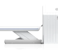
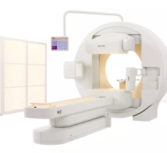
Launching at SNM, Philips new BrightView X is a scalable version of the BrightView XCT. The mid-level SPECT camera offers a cost-effective option that allows customers the flexibility to purchase SPECT now and add CT later. The transition involves a simple upgrade without a change to the room size or power requirements.
SPECT/CT has long been utilized in cardiac studies, but with Philips' upgraded BrightView XCT there is promise for applications in orthopedic studies, which is a field that typically relies heavily on X-ray imaging. At SNM 2009, Philips is showing the first SPECT/CT hybrid system to address physicians’ needs for high-quality localization and attenuation correction with a flat-panel CT, while still allowing for patient dose reduction and increased diagnostic confidence, the company said.
To improve image registration, Philips BrightView XCT uses a co-planar design that allows the acquisition of SPECT and CT with minimal table movement between scans in most cases. Reducing movement, along with the system’s wide-open gantry and large bore, contribute to improved patient comfort. Flexible breathing protocols also set it apart from other systems by allowing patients to breath normally during both SPECT and CT scans.
Along with the addition of the new Nuclear Medicine Application Suite on the Extended Brilliance Workstation, nuclear medicine is now available with the BrightView XCT; SPECT, PET and CT processing can all be accessed on one common platform, helping to speed diagnosis and streamline workflow. The BrightView XCT is a general purpose nuclear medicine SPECT/CT camera that was introduced at last year’s SNM, but is being shown with expanded applications in orthopedics at this year's SNM conference.
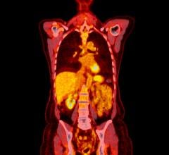
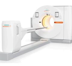
Philips is also introducing the third-generation Time-of-Flight platform for oncology on its GEMINI TF PET/CT. Time-of-flight technology measures the actual time difference between the detection of coincident gamma rays for more accurate localization, producing a higher quality image and allowing for lower tracer dose amounts in patients and shorter imaging times. According to Philips, the system is the first commercial PET/CT scanner to break the 500 Pico-second barrier, with the speed to measure a photon within seven centimeters accuracy traveling seven times around the earth in a single second.
Philips newest Time-of-Flight system, which was launched at SNM 2008, the GEMINI TF Big Bore PET/CT, is specifically designed for radiation oncologists and their patients. The Big Bore offers excellent lesion detection for diagnosis and staging with Philips revolutionary TruFlight Time-of-Flight PET technology. The system has been installed at three sites throughout the U.S., including the first installation at a proton therapy center at the University of Pennsylvania.
For more information: www.philips.com/newscenter


 July 02, 2024
