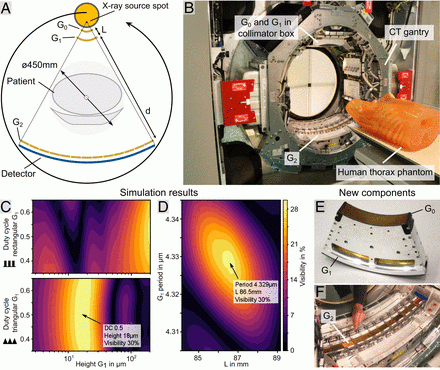
Design of the human-scale dark-field CT system. (A) Layout of the Talbot–Lau interferometer integrated into a conventional medical CT system. Bent gratings in an inverse geometry allow positioning of G0 and G1 close to the source. A large G2 is positioned close to the detector. Contrast formation is illustrated in SI Appendix, Fig. S1. (B) CT gantry equipped with a Talbot–Lau interferometer. The large G2 covering the detector is visible, while the G0 and G1 fixture is concealed by the collimator box. A human chest phantom is positioned on the patient couch. (C) Parameter analysis for rectangular and triangular G1 gratings. A maximum performance (i.e., fringe visibility) is expected for a duty cycle of 0.5 and 18.5μm18.5 μm in height for a triangular profile. (D) Simulation shows that performance is highly parameter dependent. Small deviations in G1 periodicity in the nanometer range or in length L in the millimeter range cause irreversible performance loss. (E) A specialized G0 and G1 fixture to bend gratings to focus into the X-ray source spot. Rigid mounting is important to ensure stability during continuous rotation at high centrifugal forces. (F) Stitched G2 using a modular adjustment frame to individually position a total of 13 tiles. Fine position and rotation manipulation as well as long-time stability during rotation are key aspects of this component (SI Appendix, Fig. S2). Image courtesy of PNAS
February 9, 2022 — For the first time, a team of researchers at the Technical University of Munich (TUM) has integrated the dark-field X-ray method into a CT scanner suitable for clinical use. Dark-field imaging provides additional information to conventional X-ray imaging. With the new prototype, it is now possible to produce three-dimensional dark-field X-ray images.
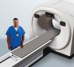
Computed tomography (CT) is one of the most important clinical methods for precise and fast diagnostics. By combining multiple X-ray images three-dimensional images of the patient are generated. With dark-field imaging now additional information on fine tissue structures, in particular in the lung, is accessible. Until now, technical challenges have prevented the integration of this new technology into clinical CT scanners to examine patients.
A team of researchers working with Franz Pfeiffer, Professor for Biomedical Physics and Director of the Munich Institute of Biomedical Engineering at TUM, has now developed a CT scanner that combines both X-ray technologies.
“For the first time, we showed that dark-field X-ray technology can also be integrated into a clinical CT scanner. Although this technology is in its early stages, pre-clinical studies with mice have demonstrated clear benefits from dark-field CT scans, especially for capturing images of lung tissue,” says Franz Pfeiffer, who headed the study. The new CT prototype has already been used successfully with a thorax phantom, a model of a human upper body, and is large enough for the intended applications with real patients.
Conventional X-ray imaging
With conventional X-ray equipment, the X-rays are attenuated by the intervening tissue as they travel from the source to the detector. This effect is used to produce images based on the varying degrees of attenuation associated with different tissue types and structures. That is why bones and similar structures, which have a stronger attenuating effect, appear white in X-rays, while more transparent tissue types such as the lung produce darker images.
Dark-field X-ray imaging
Dark-field imaging, by contrast, makes use of the small-angle scattering of the X-rays. When the X-rays interact with materials of different densities such as the interface between lung tissue and air, they are scattered. The analysis of this scattering effect yields additional information on very fine tissue structures, which is otherwise not accessible with conventional X-ray images.
Grating technology for dark-field imaging
To detect the scattering of the X-ray radiation, a set of three optical gratings is required. They are placed between the X-ray source and detector. When X-rays pass through these gratings, a characteristic pattern is produced at the detector. When a sample or person is then positioned in the beam path, this characteristic pattern is changed. These deviations are then used to analyze the structure of the sample or the person’s tissue.
New hardware and software for dark-field CT
The implementation of the dark-field method in a human-size CT scanner poses various technical challenges. Until now, this has limited dark-field CT devices to a scale much smaller than would be needed for human patients. Apart from the size, the fast rotation of the scan unit also creates special difficulties for the technical design.
The scanning unit of CT scanners, known as the gantry, rotates at very high speeds. This causes vibrations that affect the finely-tuned components in the interior of the device. Based on a detailed analysis of these vibrations, the team was able to use them to implement the required shift between the gratings needed for dark-field imaging. To analyze the scans, they developed new algorithms to filter out the vibration effects based on reference scans.
Additional information for clinical diagnostics
“With the dark-field CT prototype, we can capture conventional and dark-field X-ray images in a single scan. This yields additional information that could be used in the future not only to diagnose lung diseases, but also to differentiate between various types of kidney stones and tissue deposits,” said Manuel Viermetz, one of the two first authors of the study. As the next step, the researchers plan to further optimize the dark-field CT prototype and prepare for the first scans of human patients.
For more information: https://www.tum.de/en/

 March 19, 2025