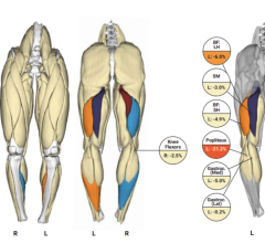
Still image illustrating the lack of lymphatic flow in the lower leg of a subject with lymphedema. Credit: John Rasmussen
June 5, 2014 — A team of researchers at the University of Texas Health Science Center at Houston (UTHealth) Medical School has developed a new technology that can noninvasively image the human lymphatic system (also known as “lymphatics”). A fluorescent dye and commercially available laser diode and military-grade night vision devices were used to visualize the lymphatic capillaries.
The human lymphatic system is an important but poorly understood circulatory system consisting of tiny vessels spread throughout the body. This “drainage” network helps guard against infections and prevents swelling, which occasionally happens when disease or trauma interrupts normal lymphatic function. Chronic swelling is the hallmark of a painful, incurable condition known as lymphedema, which often occurs after cancer therapy and can leave the limbs and other body parts disfigured for life. Detecting lymphedema early, before swelling occurs, would lead to better outcomes for patients, but the major barrier preventing early diagnosis is the lack of high-resolution imaging techniques that can resolve these tiny vessels.
Clinically, the UTHealth researchers’ device promises improvements in patient care because it allows even tiny lymph vessels to be imaged, and it can quantitatively measure fluid flow throughout the lymphatic system — two types of measurements that are impossible with current technology.
At Conference on Lasers and Electro-Optics (CLEO) 2014, to be held June 8-13 in San Jose, Calif., UTHealth scientist John Rasmussen, Ph.D., will describe how they have taken this technology, which they call near-infrared fluorescence lymphatic imaging (NIRFLI), from bench-top development to various clinical applications. "We feel that the ability to see the lymphatics will provide opportunities to revolutionize lymphatic care," Rasmussen said.
The major problem with lymphatic imaging is that the small lymphatic vessels are filled with lymph, a clear liquid that lacks the natural contrast needed to show up on modalities like computer tomography (CT) or magnetic resonance imaging (MRI). While one might think about injecting dyes or other contrast "agents" into the lymphatic vessels to make them more visible, the vessels are very difficult to find and are most often too small to insert a needle.
An existing technology, called lymphoscintigraphy, can take images of the lymphatic system following injection of a radioactive compound into or below the skin. However, lymphoscintigraphy typically takes 20 to 45 minutes to acquire a single grainy picture, and can only image the largest lymphatic vessels or trunks. The smaller vessels, which make up the bulk of the lymphatic system, are invisible to lymphoscintigraphy. In addition because of the long acquisition times, it cannot capture the real-time flow of fluid in the system.
At CLEO: 2014, Rasmussen will describe how NIRFLI allows lymphatic structures and flow to be measured quantitatively, including in the fine vessels — an improvement over lymphoscintigraphy. In addition, NIRFLI will be safer and less expensive than existing technology.
To acquire images of the lymphatics, NIRFLI uses indocyanine green dye, which is injected in tiny amounts into the skin of a patient. The dye is absorbed into the lymphatics and when illuminated by the laser diode, it emits a fluorescent light, which the device amplifies with a military-grade image intensifier (the main component in night vision goggles) and then captures with a commercial CCD digital camera.
The image intensifier enables the small lymphatic vessels to be visualized, and by taking sequences of such images, they can produce movies showing flow within the lymphatics. Rasmussen said that the most immediate promise of NIRFLI will be to diagnose and monitor the treatment of lymphedema and may also help surgeons identify and remove lymph nodes into which cancer tumors drain.
"From these images and movies, we can identify abnormal lymphatic structure and function in a variety of diseases and disorders in which the lymphatics play a role," Rasmussen said. "I think we have barely scratched the surface of what is possible."
NIRFLI was developed with funding from the U.S. National Institutes of Health and in collaboration with Eva Sevick-Muraca, Ph.D., and other scientists and engineers at UTHealth. Rasmussen added that UTHealth has licensed the technology to NIRF Imaging Inc. for commercialization.
For more information: www.cleoconference.org
 August 06, 2024