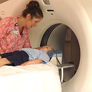
April 5, 2011 – Computed tomography (CT) examinations of children in hospital emergency departments increased substantially from 1995 to 2008, according to a new study published online and in the June print edition of Radiology. Researchers said the findings underscore the need for collaboration among medical professionals to ensure that pediatric CT is appropriately ordered, performed and interpreted.
“We need to think creatively about how to partner with each other, with ordering clinicians and with CT manufacturers to ensure that all children are scanned only when it is appropriate and with appropriate techniques,” said the study’s lead author, David B. Larson, M.D., MBA, director of quality improvement in the department of radiology at Cincinnati Children’s Hospital Medical Center in Ohio.
Advancements in CT technology like helical scanning have made it a vital tool for rapid diagnostic evaluation of children in the emergency department. Decreased scan times are especially helpful in eliminating the need for sedation in many pediatric cases.
However, the relatively higher radiation doses associated with CT, compared to most other imaging exams, have raised concerns over an increase in risks associated with ionizing radiation. A child’s organs are more sensitive to the effects of radiation than those of an adult, and they have a longer remaining life expectancy in which cancer may potentially form. In addition, the current prevalence of CT makes it more likely that children will receive a higher cumulative lifetime dose of medically related radiation than those who are currently adults.
To study CT utilization trends in children, Larson and colleagues analyzed National Hospital Ambulatory Medical Care Survey data from 1995 to 2008.
The number of pediatric emergency department visits that included a CT examination increased five-fold over the study period, from roughly 330,000 to 1.65 million, with a compound annual growth rate of 14.3 percent. The leading complaints among those receiving CT included head injury, abdominal pain and headache. The rate of imaging for abdominal pain increased the most, owing to improvements in the technology.
“We found that abdominal CT imaging went from almost never being used in 1995 to being used in 15 percent to 21 percent of visits in the last four years of the study,” Larson said. “In 1995, abdominal CT took much longer, the resolution was not as good and the research hadn’t been done to support it. By 2008, helical scanning had helped make CT very useful for abdominal imaging. It’s widely available, it’s fast and there are a lot of great reasons to do it, but it does carry a higher radiation dose.”
Larson pointed out that abdominal CT’s effective dose of radiation is up to seven times that of a head CT, suggesting that the radiation dose to children in emergency departments increased at an even higher rate from 1995 to 2008 than the rate of increase in the percentage of visits in which CT was performed.
Non-pediatric focused emergency departments made up 89.4 percent of emergency department visits associated with CT in children and increased from 316,133 examinations to 1,438,413 over the study period. Larson noted that most of the radiologists who oversaw and interpreted these studies likely were not subspecialty-trained in pediatric radiology.
“The performance of CT in children requires special oversight, especially in regards to the selection of size-based CT scan parameters and sedation techniques,” he said. “It is important to consistently tailor CT technique to the body size of the pediatric patient,” he said.
For more information: www.rsna.org


 August 09, 2024


