At the annual meeting of the AHRA, Agfa Healthcare demonstrated a full-scale model of its DR 800, presenting the unit as a "game changer" for its multifunctionality.
Diversity was on display at the Association for Medical Imaging Management (AHRA) 2019 meeting — diversity in the practice of radiology and in product portfolios; diversity that translates into the marketplaces as complexity and uncertainty.
In July, show-goers crammed a symposium titled “Imaging Market Outlook at the AHRA 2019” where Stuart Clark, managing director and national spokesperson, The Advisory Board, described the conflicting and contrary factors that could roil the U.S. marketplace over the coming years.
A Diversity of Forces
Beginning with an analogy that contrasted airlines before and after deregulation, Clark explained how conflicting forces will disrupt an imaging marketplace. The real challenge will be recognizing how these forces affect and influence each other.
Utilization of medical imaging services in outpatient environments could rise, Clark said, but so might their charges. Rising prices could lead to efforts to control costs, steer patients to lower cost services and to develop new regulations. The uncertainty was as disquieting as the interaction of forces was complex. The years ahead would be marked in the imaging marketplace by a messy process, he predicted, one marked by disruptions.
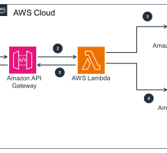
One of the unknowns that could affect the U.S. imaging marketplace is the degree to which artificial intelligence will be adopted — and how its use will affect the imaging marketplace. Speaking July 21 at AHRA 2019, Siemens Healthineers executives Wesley Gilson and Peter Shen described in their session AI’s “implications for advanced imaging and precision medicine.” Before the meeting, Gilson described how the company is leveraging AI to make its radiology equipment generate results faster and more reproducibly, potentially boosting productivity by speeding up clinical workflow, preventing diagnostic errors, even reducing missed
billing opportunities.
Spotting Lies And Liars
Traci Brown packed a session titled “Liar, Liar Pants On Fire.” Everyone lies, Brown explained. But there are times when identifying lies can save radiology administrators “time, money and energy.” She illustrated that point with stories of technologists who said they were not the ones who dropped (and broke) X-ray detectors. Or who took time off to attend the funeral of their fathers (who had already died).
Brown explained that spotting lies requires paying attention to the person’s body language — the head bobbing “yes” when the voice is saying “no;” feet shuffling as if the speaker wants to run away; lips that curl inward as if to keep facts from spilling out a moment later; eyes rapidly shifting up, down and sideways; outrage rather than quiet indignation.
“Body language informs the conversation,” she said. “It tells you where to dig — what questions to ask and where to go.”
Embodiments of Pragmatism
Citing the need to control soaring costs, payers continue trying to reform healthcare. The shift from volume- to value-based imaging is one of these efforts. Determining the value of imaging is not easy, but radiology practices need to be engaged in the value paradigm, according to John Carrino, M.D., in a July 21 session titled “Challenges and Opportunities for Radiology to Prove Value in Alternative Payment Models.” Focusing on value “doesn’t mean that you buy into all these models,” Carrino told Imaging Technology News (ITN) after his presentation. “But you need to be aware of (alternate payment models) and have a strategy that works for your practice.”
Effectively managing risk in magnetic resonance imaging (MRI) can save money by increasing efficiency. And it can benefit the patient, said John Karis, M.D., director of neuroradiology at the Barrow Neurological Institute. Speaking on the topic July 21, Karis told ITN before the meeting that an early step in the process is to make “intelligently formed decisions … about what would be the best agent to use.”
Macrocyclic contrast agents have the best safety profile of all commercially available MR media, Karis said. So compelling is their safety profile that the Phoenix, Ariz.-based institute switched to using macrocyclics for the vast majority of MR contrast-enhanced scans. It was out of concern for patient safety that the switch was made, he said. Although these agents are more expensive than linear ones, their small cost differential can be offset by improved efficiency, according to Karis.
Innovation is trending toward improved efficiency — but not at the expense of patient safety, according to Michael Perez de Utrera. The diagnostic imaging manager of the seven Leon Medical Centers in Miami, Fla., spoke on the subject July 24 at AHRA 2019, explaining that although reimbursement rates continue to lessen, “patient safety always comes first.”
Smart devices can help achieve both objectives, he said, noting how the integration of smart devices with electronic medical record systems “definitely improves efficiency.” This is exemplified at the Leon Medical Centers by the use of software that helps manage contrast and patient radiation exposure.
Venturing Into CT
When providers develop their own imaging protocols, they are wasting time and money, according to Timothy Szczykutowicz, Ph.D. Sites that do this are “re-inventing the wheel,” said the associate professor in the University of Wisconsin Departments of Radiology, Medical Physics and Biomedical Engineering. Instead they should adopt protocols that already exist.
About 1,500 sites around the world have obtained UW protocols for computed tomography (CT) under license with GE Healthcare, according to Szczykutowicz, an expert in CT protocol management and optimization who co-chairs the task group on protocol management at the American Association of Physics in Medicine. He and colleagues addressed the value of protocol standardization
July 23 at the AHRA annual meeting.
Speaking with ITN, Szczykutowicz noted that the licensed protocols, which address musculoskeletal, chest, pediatrics, cardiovascular and brain CT, are specific to GE CT scanners.
A big trend in the cardiac application of CT has been the use of noninvasive fractional flow reserve CT (FFR-CT). This application can in some circumstances reduce the need for conventional FFR in the cath lab, according to evidence presented July 22. Session presenters noted in a slide that the CT-based procedure can be used to identify the patients most likely to test positively for coronary artery disease.
FFR-CT has been proven to reduce unnecessary hospital admissions, they said in their presentation, citing research showing that the procedure provides the information that cardiologists need without the expense, time or patient inconvenience of tests done in the nuclear medicine or cardiac catheterization labs.
On the Exhibit Floor
Some exhibitors strummed broad-based, yet familiar themes; others sounded off about issues closing in or already in play. The vast majority paid homage to the staples of medical imaging — X-ray and ultrasound.
Canon Medical Systems welcomed floor-walkers with a booth that depicted the diversity in the company’s portfolio. At one end of the booth was portable X-ray and flat panel detectors; at the other was portable ultrasound; in between were desktop monitors featuring enterprise imaging from Canon’s Vital Images. Monitors scattered around the booth displayed images of advanced products previously sold under the corporate umbrella of Toshiba Medical Systems, which Canon formally acquired in early 2018.
Nearby was Shimadzu Medical Systems, which broadened its appeal as a digital radiography (DR) neutral vendor by featuring Smart Stitch (from Canon), a long-imaging capability for spines and legs, and Dynamic Digital Radiography (DDR), a recently FDA-cleared capability that visualizes movement using conventional X-ray. Shimadzu collaborated with Konica Minolta in the development of DDR, combining Konica’s advanced image processing with its own RADspeed Pro X-ray imaging system. Shimadzu also framed its FDA-pending FLUOROspeed X1, a radiography/fluoroscopy (RF) table system, for supporting advanced RF features on a conventional table.
On the exhibit floor, Konica Minolta Healthcare Americas highlighted DDR as demonstrating its commitment to innovation and cost effectiveness, as well as its ultra U-arm and data analytics exemplifying efficiency and cost effectiveness. The overall pitch was to administrators who have to do more without commensurate budgets; affordability for rural as well as big city medical facilities; and technology that helps radiologists and staff make “better decisions sooner.”
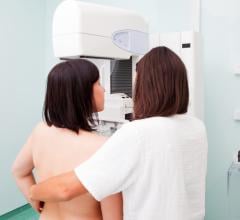
At AHRA 2019, Fujifilm Medical Systems USA sponsored a symposium on long-length DR. Gregg R. Cretella, the company’s national director of clinical operations, told ITN prior to the meeting that DR long-length detector technology and image processing algorithms are changing the utilization of this modality in spine imaging. On the exhibit floor Fujifilm showcased mammography developments designed to improve image quality and lower patient dose.
Focused on women’s health, Hologic highlighted its Unifi Analytics, a business intelligence tool designed for the mammography enterprise. The objectives behind its use are to maximize efficiency and reduce the downtime of mammography equipment, at least partly by predicting equipment failures so administrators can plan for repairs.
Siemens Healthineers showcased Mammomat technologies as exemplifying the company’s theme of digitalization as a means for developing patient insights. GE Healthcare featured wireless detectors with 100 micron resolution and high detective quantum efficiency (DQE) compatible with the company’s portable X-ray machines, which were shown on the booth. Philips Healthcare illustrated its DigitalDiagnost C90 using a desktop model that highlighted tableside controls and an optical camera (built into the overhead tube crane) to help in patient positioning.
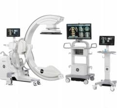
In its booth, Agfa Healthcare demonstrated a full-scale model of its DR 800. Billed as a “game changer” for its multifunctionality, the DR 800 offers radiography, fluoroscopy and advanced clinical applications. Agfa’s Musica algorithms are built in for image processing and workflow. Through a partnership with Hitachi, the DR 800 is sold mainly to rural and community facilities. Agfa sells the unit primarily to larger medical facilities.
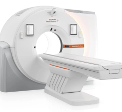
At the meeting, Hitachi Healthcare America focused on rural and community sites, emphasizing patient comfort with wide bore CT and MRI, while talking up the flexibility of ultrasound scanners. Still finalizing an agreement to sell its IT business to Philips, Carestream Health emphasized X-ray equipment, including portables and a cone beam CT for extremities. Avoiding the expense of setup and tear down, Del Medical relied on pictures to pitch its rad rooms.
Samsung framed its X-ray products as being entirely company-made — from detectors to software, X-ray portables to the rad room with its overhead tube crane and floor-mounted wall stand. Detectors, including the newly introduced iQuia, are optimized to deliver low-dose radiation. Neusoft presented its products as inexpensive and delivering high performance, exemplified by the company’s 64-slice CT, which comes equipped to perform cardiac calcium scoring.
With a 25-year history in medical imaging, Sectra framed itself as an IT software company with the knowledge and experience to bring together disparate elements of consolidated enterprises as well as those still in the process of consolidating. Fully and securely image enabling electronic medical record systems and “basically any ‘ology’ that can acquire images” was the pitch.
Novarad unveiled a system for sharing images electronically with its CryptoPACS (crypto patient archive and communications system) designed to replace CD image sharing. Already for this purpose, Ambra Health seamlessly connects facilities. IT provider Paxera Health claimed the ability to “do it all” from enterprise imaging to PACS to sharing patent images electronically. Precision Image Analysis (PIA) promoted off-site image post-processing as a way providers could conduct more exams.
Imalogix framed itself as a vendor-agnostic information service that helps customers run efficiently. Optimizing dose is important, booth staff agreed, but so is identifying ways to improve image quality and exam consistency.
ScreenPoint Medical demonstrated 2-D and 3-D versions of an artificial intelligence-based system for interpreting digital mammograms. The 3-D version, called Transpara, has the CE mark for European Union sale but is not yet cleared by the FDA for sale in the U.S. Transpara assigns numbers to images that reflect the likelihood of cancer — the higher the number, the more likely the cancer.
Bracco Diagnostics and Bayer featured proprietary lines of power injectors, as well as software for safely and easily administering contrast agents. In one configuration, the companies’ different software help manage the administration of contrast media.
In another, their software packages track patient radiation exposure.
Kopp Development showed how far ferromagnetic detectors have come. Built into the doorways of MR suites, Ferralert Encompass automatically detects and gives the approximate location on the person of ferromagnetic objects passing through the doorway. Alternatively, as shown in other booths, such as shielding provider ETS Lindgren, the underlying technology can be configured to scan people getting ready to enter the MR suite.
Greg Freiherr is a consulting editor to Imaging Technology News (ITN). Over the past three decades, he has served as business and technology editor for publications in medical imaging, as well as consulted for vendors, professional organizations, academia and financial institutions.

 August 06, 2024