ScImage announced the immediate availability and deployment of updated quantification metrics based on guidelines published jointly in January 2015 by the American Society of Echocardiography (ASE) and the European Association of Cardiovascular Imaging (EACVI).
In 1934, a Senate committee opened hearings its chairman said would show that America’s involvement in WWI was “not a matter of national honor and national defense, but a matter of profit for the few.” Ninety-three hearings and 200 witnesses, however, did little to support that claim. Rather the hearings drew increased attention to arms manufacturers as “merchants of death” and inspired Congress to pass three neutrality acts that signaled “profound American opposition to overseas involvement” in the years preceding America’s entry into WWII.[1]
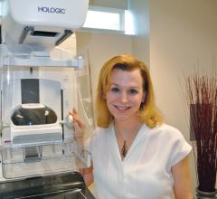
Despite its small size, Gritman Medical Center, a 25-bed hospital in Moscow, Idaho, has a reputation for thinking big, especially when it comes to women’s health. That’s why in May 2014 the hospital became the very first facility in the region to install a Hologic 3D mammography system and began to offer breast tomosynthesis services to its patients. The addition of the Hologic system represents a significant capital investment for the hospital, especially since they decided from the start not to charge women an additional fee for 3D mammograms. “3D was not a financial decision, it was a decision based upon what is the right thing to do for the women we serve,” noted Christin Reisenauer, M.D., director of imaging at the hospital’s Patricia J. Kempthorne Women’s Imaging Center. “We knew we were missing breast cancers with 2D mammography, cancers potentially visible with breast tomosynthesis, and that just wasn’t fair to the women of our area.”
While most women understand the importance of health screenings, an estimated 72 million have missed or postponed a ...
Breast density may be one of the strongest predictors of the failure of mammography to detect cancer, according to an educational session presented at the 2014 Radiological Society of North America (RSNA) conference in December. The topic of breast density was a prominent one at the meeting, and many healthcare providers are beginning to look beyond just using traditional mammography to assess whether or not a woman has breast cancer. The push for additional screening is becoming prevalent, and many states are enacting laws that require women to be notified if they have dense breast tissue and what that means in terms of the ability to accurately find cancer.
It’s no surprise to state that healthcare technology has been slow to mature. However, much of this perception has come from lack of a greater picture; the proverbial “forest for the trees.” No longer is radiology defined as a siloed infrastructure or walled-off service line catering only to physicians utilizing the platform, but it is now considered the transactional hub of services driving a consumer-based healthcare product. Although reimbursement and organizational strategies have driven much of the recent evolution, consumer awareness and attention to healthcare costs are pushing a demand for greater visibility into the mechanisms of healthcare.

SPONSORED CONTENT — Fujifilm’s latest CT technology brings exceptional image quality to a compact and user- and patient ...
Radiologists play a key role in the cancer care continuum after the initial diagnosis of prostate cancer. This includes regular imaging assessments to monitor growth, and tracking tumor changes to measure the effectiveness of various therapies. For the radiologist evaluating prostate cancer, it is key to first determine the severity of the disease, which determines whether the patient will be monitored over the course of several years, or if immediate treatment is required.
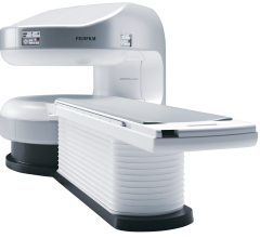
Fujifilm’s APERTO Lucent is a 0.4T mid-field, open MRI system addressing today’s capability and image quality needs ...
Heart Imaging Technologies announced the release of Precession, a cardiac magnetic resonance solution which allows viewing, analysis and reporting all within a standard web browser.
Multiple studies and products were presented at the 2014 Radiological Society of North America (RSNA) conference in December about emerging technologies in breast imaging, with a focus on how they will affect women who have dense breasts. Most researchers have been comparing the utility of mammography screening versus tomosynthesis.
Barco announced the launch of a new diagnostic display system, Nio 5MP LED. Cleared by the U.S. Food and Drug Administration (FDA) for radiology and mammography and featuring a number of image-enhancing technologies, the 5MP display provides excellent image quality for confident diagnoses.
SPONSORED CONTENT — Fujifilm’s latest CT technology brings exceptional image quality to a compact and user- and patient ...
As healthcare and radiology continue to evolve rapidly, flexibility in the operating room (OR) and imaging lab are key. Consequently, imaging systems need to be versatile as well as able to handle a variety of applications and adapt to any space. These needs have led to the increasing popularity of mobile C-arms, the latest generation of X-ray technology. Today, they are being adapted to handle fluoroscopy, vascular imaging, cardiology and 3-D imaging.
Recently, the Centers for Medicare and Medicaid Services (CMS) issued a final national coverage determination, effective immediately, that provides for Medicare coverage of screening for lung cancer with low dose computed tomography (LDCT). This screening gives at-risk seniors unprecedented access to care.
Cancer patients with limited brain metastases (one to four tumors) 50 years old and younger should receive stereotactic radiosurgery (SRS) without whole brain radiation therapy (WBRT), according to a study in the March 15, 2015 issue of the International Journal of Radiation Oncology • Biology • Physics (Red Journal).
SPONSORED CONTENT — EnsightTM 2.0 is the newest version of Enlitic’s data standardization software framework. Ensight is ...
Accuray Inc. announced that the first patient treatment has been completed using the CyberKnife M6 System with the InCise Multileaf Collimator (MLC). The treatment was administered as a multidisciplinary effort between Steven Burton, M.D., from the Department of Radiation Oncology, and Johnathan Engh, M.D., from the Department of Neurosurgery at the University of Pittsburgh Medical Center (UPMC) in Pittsburgh, Pennsylvania.
New and updated American College of Radiology (ACR) Appropriateness Criteria now help healthcare providers choose the most appropriate medical imaging exam or radiation therapy for more than 1,000 clinical indications. These continually updated criteria are a national standard developed by expert panels of physicians from many different medical specialties.

SPONSORED CONTENT — EnsightTM 2.0 is the newest version of Enlitic’s data standardization software framework. Ensight is ...
CompView Medical (CVM) introduced an all-in-one equipment manager, visualization and ergonomic boom system — the NuCart.
Frequent use of lead aprons in the interventional lab and radiology departments to protect against radiation exposure is associated with increased musculoskeletal pain, according to a study published in the Journal of the American College of Cardiology.
Disparities in the level of awareness and knowledge of breast density exist among U.S. women, according to the results of a Mayo Clinic study published in the Journal of Clinical Oncology.
Medical imaging plays a vital role within the healthcare enterprise, both from a value and patient care perspective. However, a significant number of radiology departments across the U.S. are operating with systems that are limiting their true potential. In order to prosper, enterprises require next-generation, enterprise-wide medical imaging solutions with proven capabilities to enhance performance in multi-site, large-scale, consolidation-friendly environments.
University of Tubingen and Mediso Ltd announced at the 4th Tubingen PET/MR Workshop a collaboration to develop a whole body preclinical positron emission tomography (PET) insert based on silicon photomultiplier sensor technology. This system can be inserted into a high field (7T) preclinical magnetic resonance imaging (MRI) scanner to enable simultaneous PET/MR imaging.
GenomeDx Biosciences announced the publication of a positive validation study for the Decipher Prostate Cancer Classifier, a genomic test for prostate cancer.


 March 03, 2015


