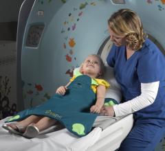
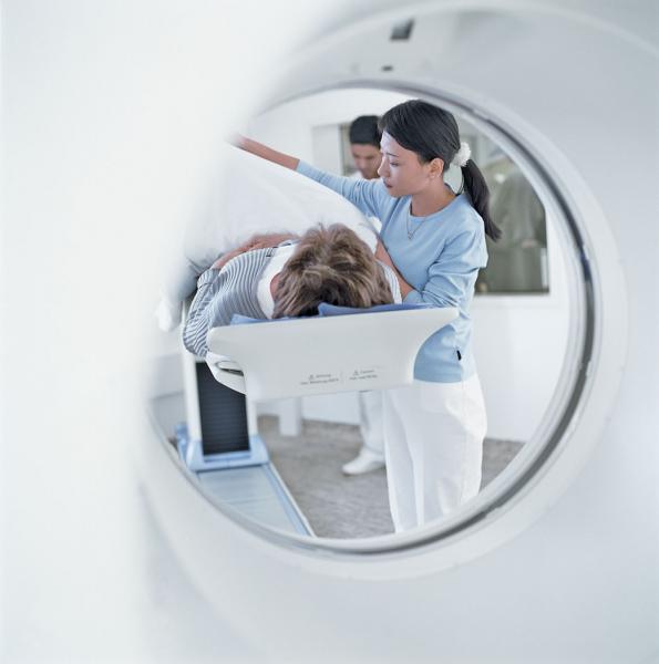
Safety accidents drive up costs, damage reputations and lower patient satisfaction scores. For instance, if a patient falls off a system’s table after being sedated and is badly injured, it can result in significant repercussions for the provider. This type of scenario is rare, but still causes concern for health systems nationwide.
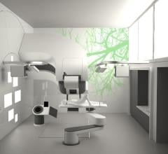
The Texas Center for Proton Therapy announced it has selected RayStation from RaySearch Laboratories as its treatment planning software. The 63,000-square-foot facility in the Dallas/Fort Worth area will have capacity to treat more than 100 patients per day in three treatment rooms when it opens in late 2015. It will be the first stand-alone LEED-Certified (Leadership in Energy and Environmental Design) proton therapy center in the United States.
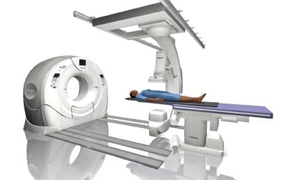
All of the major vendors in the United States introduced new systems and technologies in the past few years to reduce dose and enhance visualization in the cath lab. The vendors have tailored their systems into various models for specific specialties and at various price points depending on the degree of functionality. Most vendors also offer software to enhance visualization of stents and other devices.
While most women understand the importance of health screenings, an estimated 72 million have missed or postponed a ...
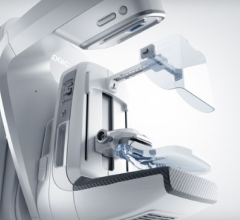
iCAD Inc. announced the launch of the iReveal breast density module as the latest addition to the company's PowerLook Advanced Mammography Platform (AMP). The iReveal software is designed to deliver automated, rapid and reproducible assessments of fibroglandular breast density to help identify patients that may experience reduced sensitivity to digital mammography due to dense breast tissue.
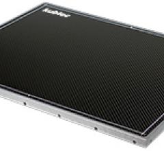
Kubtec released the new 17 x 14-inch Digiview 395 wireless flat panel detector for medical imaging. Lightweight, durable and easy to use, the Digiview 395 incorporates Kubtec's imaging capabilities and rapid image acquisition and display with wireless freedom. Designed for versatility and portability, the detector can be used anywhere medical imaging is required. It features wireless data transmission, giving medical personnel immediate access to critical information for faster interpretation and diagnosis.

SPONSORED CONTENT — Fujifilm’s latest CT technology brings exceptional image quality to a compact and user- and patient ...
London Imaging Centre has partnered with Mach7 Technologies to streamline image management and workflow through implementation of the Mach7 Enterprise Imaging Platform.
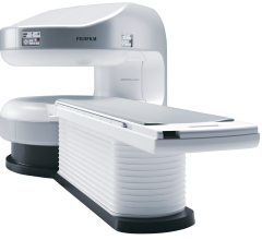
Fujifilm’s APERTO Lucent is a 0.4T mid-field, open MRI system addressing today’s capability and image quality needs ...
EOS imaging announced dual EOS imaging system installations at two of the leading, and largest, academic hospitals in Germany – Heidelberg University Orthopedic Hospital and the Medical Center of the University of Munich.
The Dr. Samadi Prostate Cancer Center in New York City is now offering a full suite of new genetic testing methods for diagnosing prostate cancer. The new methods are intended for men with an elevated prostate specific antigen (PSA) level or who have had a biopsy.
IBA (Ion Beam Applications S.A.) has signed three separate binding term sheets with Proton Partners International (PPI) to install three of its Proteus One compact proton therapy solutions in private clinics in the United Kingdom. The agreements contain significant and immediate down-payments.
SPONSORED CONTENT — Fujifilm’s latest CT technology brings exceptional image quality to a compact and user- and patient ...
Ten weeks of intensive reading intervention for children with autism spectrum disorder (ASD) strengthened loosely connected areas of their brains that comprehend reading, University of Alabama at Birmingham researchers found. At the same time, the reading comprehension of those 13 children, whose average age was 10.9 years, also improved.
Medraysintell released its updated World Market Report and Directory on Nuclear Medicine, Edition 2015, in late June, providing a description and analysis of the latest developments in nuclear medicine. The 920-page document covers 335 radiopharmaceuticals and radionuclides and 160 companies and institutions active in nuclear medicine.
Years ago in Vegas I came face-to-face with the strain of radiology. On-site, at an imaging practice built around managed care, I sat from 9 in the morning until 7 that night observing (as unobtrusively as possible) a radiologist. At day's end he retreated to his office, dragging me with him for a recap of the day, his eyes puffy and bloodshot.
SPONSORED CONTENT — EnsightTM 2.0 is the newest version of Enlitic’s data standardization software framework. Ensight is ...
Just a few short years ago, the only options for breast imaging were 2-D mammography, but thanks to rapidly advancing technology to address accuracy concerns in reading dense breast images, today there several options, including 3-D tomosynthesis.
With radiology departments generating so much imaging data on a daily basis, and as medical imaging technology continues to advance, many existing picture archiving and communication systems (PACS) and other storage solutions simply do not have the capacity to handle multiple terabytes of data on-site. As a result, hospitals and other healthcare organizations are turning to cloud solutions. It is a buzzword that many are familiar with, but one that many still do not fully understand.
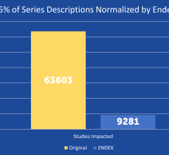
SPONSORED CONTENT — EnsightTM 2.0 is the newest version of Enlitic’s data standardization software framework. Ensight is ...
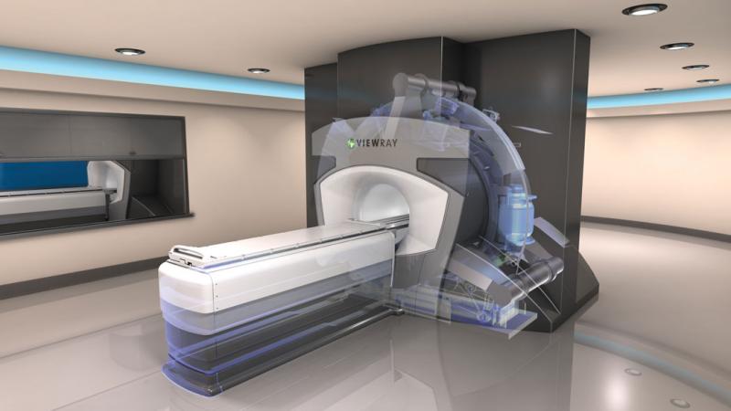
Image-guided radiation therapy (IGRT) affords greater accuracy of dose distribution during cancer treatment, allowing the radiation oncologist to see how a tumor is responding over the course of treatment. This has traditionally been accomplished with computed tomography (CT) or X-ray scans, which requires extra radiation exposure for the patient, with relatively poor contrast in soft tissue due to uniform electron density. Since treatment is only as good as the images provided, efforts are under way to find a better modality — and magnetic resonance imaging (MRI) may hold the answer.
Molecular imaging is a broad and dynamic field that encompasses a range of image technologies that allow physicians and researchers to noninvasively visualize biological processes at the cellular and molecular level. Currently, the vast majority of clinical applications of molecular imaging use radiolabeled compounds (radiopharmaceuticals) that are detected with gamma cameras, single-photon emission computed tomography (SPECT) or positron emission tomography (PET), depending on the type of radioactivity used. Molecular imaging techniques typically complement more anatomic-based imaging modalities such as computed tomography (CT) and magnetic resonance imaging (MRI), and hybrid imaging modalities including SPECT/CT, PET/CT and more recently PET/MRI are available for clinical use. Together, multimodality molecular imaging can more accurately localize and characterize disease processes than either modality alone.
State-of-the-art imaging displays are a must for the healthcare arena, and today have an increasing capacity to offer large, high-resolution displays, color accuracy, calibrated brightness, advanced connectivity, optimized workflow and high contrast, just to name a few. A variety of vendors continue to improve the technology to better display imaging modalities, including computed tomography (CT), magnetic resonance imaging (MRI), X-ray, positron emission tomography (PET), mammography and ultrasound, and to ensure screens remain DICOM (digital imaging and communications in medicine) compliant.
The U.S. Food and Drug Administration (FDA) announced its latest efforts in supporting the Bonn Call for Action on radiation protection.
The future of picture archiving and communication systems (PACS) is changing. What emerged more than 20 years ago with a bang and helped change the face of diagnostic imaging and productivity is now seeing its own changes as new technology advances. Functions that once were exclusive to PACS are shifting to different applications.
Governor Bobby Jindal signed the 24th density reporting bill (House Bill 186) into law. The Act, known as the Monica Landry Helo Early Detection Act, will become effective January 1, 2016.


 July 07, 2015

