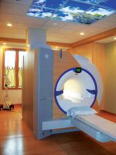
While the harmful effects of ionizing radiation have been common knowledge for some time, it is only in the last decade or so that there has been a heavy focus on patient safety in radiology. Unfortunately, this was largely because of heavily reported cases of patients suffering physical trauma due to being excessively dosed during computed tomography (CT) examinations. National organizations such as the American College of Radiology (ACR), Medical Imaging and Technology Alliance (MITA) and the Joint Commission have devised various standards related to radiation safety, but much of the progress of the last 10 years can be attributed to the efforts of individual states, which are in turn inspiring others to take action.


Much has been documented about the role of enterprise imaging as part of the larger effort to integrate a single longitudinal patient record. Despite the many ways an imaging effort reflects an organization’s electronic medical record (EMR) effort, there remain some distinct differences. For one, an enterprise imaging effort requires more careful understanding of peripheral and disparate systems. As an initiative, this primarily will serve as a support technology to the EMR and will be successful based on how it optimizes existing systems. Second, departmental workflow must remain uncompromised as part of an efficacious care delivery ecosystem. The challenges associated with deploying an EMR and the well-documented dissatisfaction on clinical workflow cannot, and should not, characterize enterprise imaging.


According to the National Institutes of Health (NIH), precision-based medicine is an emerging approach for disease treatment and prevention that takes into account individual variability in genes, environment and lifestyle for each person. This allows doctors to better tailor a treatment protocol for each individual, rather than the standard “one-size-fits-all” approach that is based on average generalizations.


According to a recent study, one in four women with breast cancer who opt for a breast-saving lumpectomy will need a second surgery — increasing both costs and the risk of complications.

Hologic, Inc. announced that the Genius 3D Mammography exam is now the only mammogram that is FDA-approved as superior to standard 2-D mammography for routine breast cancer screening of women with dense breasts.


Editor’s note: In this article, David A. Strahle, M.D., candidly shares how and why he and his team became involved with the systematic development of an abbreviated protocol for screening breast magnetic resonance imaging (MRI) and what they learned after carefully evaluating the data. This original research was conducted by Strahle, Dorothy R. Pathak, Ph.D., Arlene Sierra, M.P.A., Sukamal Saha, M.D., and Catherine Strahle, D.O., and Kiran Devisetty, M.D.


Perhaps no issue has taken on more prominence in radiology than radiation safety for both patients and hospital staff. Several regulatory bodies have enacted guidelines in recent years to improve radiation dose monitoring and reporting for computed tomography (CT), including the Medical Imaging and Technology Alliance (MITA), which created the XR-29 “Smart Dose CT” standard. Beginning Jan. 1, 2016, the standard levied a reduction in Medicare reimbursement on the technical component of all diagnostic CT exams conducted on a non-compliant scanner. The initial reduction was set at 5 percent, but it jumped to 15 percent as of Jan. 1, 2017, giving providers and vendors even more incentive to closely monitor and manage dose.


Healthcare is now the most cyber attacked industry. According to a white paper published by Healthcare IT News with information provided by Avaya Inc., 89 percent of healthcare organizations have experienced a data breach involving the loss or theft of patient data, costing the industry $6.2 billion. According to the paper, the average cost of each hack was $2.2 million.  

Subscribe Now