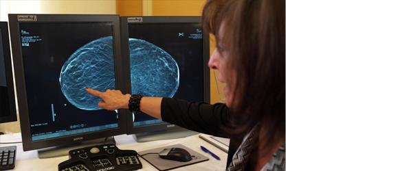
April 7, 2010 - A dual-modality tomographic (DMT) breast scanner, developed by researchers at the University of Virginia Health System (UVA), in a pilot study pinpointed to a much finer degree than mammography, ultrasound, MRI and even a needle biopsy, the location of breast masses and more accurately distinguished between cancerous and harmless lesions.
The pilot clinical study, led by Mark B. Williams, Ph.D., associate professor of radiology, biomedical engineering and physics at the University of Virginia, appears in the April 2010 issue of Radiology.
The DMT breast scanner works by coupling 3D digital X-ray breast tomosynthesis with 3D molecular breast imaging tomosynthesis, a technique that uses intravenously injected compounds (tracers) that are absorbed to a much greater degree by malignant lesions than benign ones. A special camera positioned in close proximity to the breast then performs functional imaging of the entire breast, while the digital X-ray tomosynthesis obtains co-registered structural images. The device runs the scans sequentially, obtaining both types of images with the breast in the same, immobilized position.
According to the researchers, the scanner particularly useful for women with radiographically dense breasts. Williams points out, this pilot clinical study of the 17 women tested could have far greater implications.
For more information: www.healthsystem.virginia.edu


 August 06, 2024


