Women may sometimes choose a mastectomy over radiation therapy because either they cannot meet the daily demands of a five to seven-week radiation treatment schedule or they fear recurrence and feel a mastectomy is a safer choice.
However, a recent study presented at the 2009 American Society of Clinical Oncology found that partial-breast irradiation (PBI) offers the same reduction in recurrence as whole-breast irradiation.
The hope is that recent advances in PBI, such as intraoperative radiotherapy (IORT), intracavitary brachytherapy (MammoSite), or 3D conformal/external beam radiotherapy, will demonstrate both that PBI reduces recurrence after lumpectomy and shortens radiation treatment length.
Partial vs. Whole-Breast Irradiation
Can PBI match up to whole-breast irradiation? Many investigators are searching for the answer to that question by testing new technologies and approaches to delivering this type of irradiation.
In a study presented at the 2009 American Society of Clinical Oncology Annual Meeting, researchers found that partial breast irradiation does not jeopardize survival and may be used as an alternative to whole breast radiation.1
The purpose of the study was to compare treatment outcomes in patients with breast cancer treated with PBI and of those treated with whole-breast radiation therapy. The primary outcome was overall survival and secondary outcomes were locoregional, distant and supraclavicular recurrences.
Dr. A. Valachis, lead investigator, and the team conducted a systematic review of published meta-analysis of randomized clinical trials comparing PBI versus whole breast radiation therapy. They identified three trials with a pooled total of 1,140 patients and found there was no statistically significant difference between partial and whole breast radiation arms associated with, distant metastasis or supraclavicular recurrences. However, PBI was statistically significantly associated with an increased risk of both local and regional disease recurrences compared with whole breast radiation.
They concluded: “partial breast irradiation does not jeopardize survival and may be used as an alternative to whole breast radiation. Nevertheless, the issue of locoregional recurrence needs to be further addressed.”
Intracavitary Brachytherapy
One of the leading PBI solutions that holds promise when compared to whole-breast irradiation is the MammoSite 5-Day Targeted Radiation Therapy, which is breast brachytherapy for treatment of breast cancer.
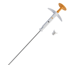
A MammoSite catheter with a balloon at the tip is temporarily implanted into the breast zone where the tumor was removed. After the lumpectomy radioactive seeds are fed through the catheter and are temporarily implanted into the tumor site. The benefit here is that the radioactive seeds are directly and immediately placed into the lumpectomy cavity. Brachytherapy radiation treatment can be completed in as little as five days, whereas traditional radiation therapy after a lumpectomy can take as long as seven weeks. The rapid completion of brachytherapy can also enable patient to receive chemotherapy sooner, if necessary.
“The clinical advantages of delivering treatment with MammoSite on a five-day radiation therapy schedule, include: targeted radiation, which limits healthy tissue from exposure; there are no side effects associated with whole breast radiation; and it’s completed in a shorter period of time; five days versus six to seven weeks,” said Greg Amante, marketing manager, MammoSite.
While there is no clinical data demonstrating that the MammoSite system is more efficacious than external beam radiation, Amante explains, “It is not more efficacious but equivalent to whole breast external radiation therapy at the five-year mark from a recurrence rate standpoint.”
In a study to determine whether MammoSite provides effective treatment for women with early-stage disease, “Partial-Breast Irradiation Therapy With MammoSite Appears to Offer Similar Results as Whole-Breast Irradiation Therapy,”2 Dr. F. Vicin, et al, found that lumpectomy followed by whole-breast irradiation is as effective as mastectomy in treating women with early-stage breast cancer.
Amante added that MammoSite’s system compared to other available breast brachytherapy treatments has “the longest term outcomes data available — five years published from FDA trial; the largest compilation of patient follow-up — 1,440 women enrolled in the ASBS MammoSite registry study; and over 50,000 women have been treated since 2002.”
While balloon brachytherapy, consisting of one catheter attached to a balloon inserted through a single-entry point, is easier to perform than interstitial brachytherapy, involving 15-20 catheters individually inserted in the breast, not everyone is a candidate for it. Patients who receive balloon brachytherapy should have a seroma still present and spherical in nature. This leaves many women ineligible for treatment with the balloon.
Another approach is to use a single-entry, multicatheter applicator. The SAVI device can treat women with cavities as small as 5-10 cc.
Before SAVI, these women would have had far fewer options for breast brachytherapy and may have been forced to choose whole breast irradiation. Physicians can customize radiation based on the size and shape of the cavity to deliver radiation with precision to the cancerous site, avoiding critical structures like the skin, chest wall or lungs. This technique broadens the pool of candidates who are eligible for breast brachytherapy.
Faster Delivery of PBI
Fast is better in the case of radiation dose delivery, as long as accuracy is not compromised. With accelerated, partial breast irradiation (APBI), the goal is to shorten the treatment time, while using a less invasive, more focused treatment without compromising survival.
In fact, APBI can shorten treatment times from the six to seven weeks to between one and five days. It also narrows the treatment area from the entire breast to the area of the breast immediately around the lumpectomy site, where most cancers are likely to recur.
APBI has been used in limited trials in several hundred patients over the last 10 years. Recent trials showed that in properly selected breast cancer patients — generally women with a small tumor, clear surgical margins after a lumpectomy, and preferably no lymph nodes containing cancer — APBI worked just as well as whole breast radiotherapy. In these early studies, investigators performed interstitial brachytherapy, but newer techniques may provide the same efficacy while delivering radiation in a faster and more convenient manner.
In an IRB approved clinical trial currently underway at Stanford University Medical Center, led by Dr. Frederick Dirbas, assistant professor of surgery, and by Dr. Donald Goffinet, professor of radiation oncology, investigators are using three new approaches for APBI.3 These three approaches are: intraoperative radiotherapy (IORT), which takes one day, intracavitary brachytherapy (MammoSite), a five-day process, and 3D conformal/external beam radiotherapy, also lasting five days.
Intraoperative Radiotherapy
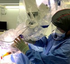
Intraoperative radiotherapy (IORT) is one of three approaches used for accelerated, partial breast irradiation at Stanford, as the treatment potentially reduces the time required for radiation therapy by delivering it immediately during surgery before any remaining cancer cells have a chance to grow.
A new technology from Xoft Inc. uses a skin and surface treatment applicator with the Axxent Electronic Brachytherapy (eBx) system to deliver surface brachytherapy, including intraoperative radiation therapy (IORT). IORT, utilizing the Axxent system’s miniaturized X-ray source that can deliver localized and targeted radiation treatment, is designed to minimize radiation exposure to surrounding healthy tissue and enables physicians and treating professionals to remain in the operating room with the patient.
“The Axxent is 510(k) cleared as a surface applicator for intraoperativer breast lumpectomy. It is used for delivering a single shot of radiation in the surface for skin cancer that is shallow, well-defined and less 5 cm in diameter,” explained Randall W. Holt, Ph.D., clinical director of medical physics for Xoft.
One of the advantages of intraoperative treatment over traditional brachytherapy is that it potentially reduces the time required for radiation therapy by delivering it immediately during surgery before any remaining cancer cells have a chance to grow.
“The long-term goal is to show it’s more effective than five-day treatment regimen compared to standard boost. In cases of previous radiation and recurrent cancer, where there is no further option, this could be effective,” said Holt.
According to Holt, in a current study in Europe, doctors are using similar technology on 1,200 patients. The protocol in the study is to use the intraoperative treatment instead of using a follow-up boost. “The standard protocol is to give 20 Gy to the surface of balloon, then five weeks of postoperative boosts. Early results show the IORT method appears to be somewhat better in terms of patient outcomesfive-year recurrence rate 4.2 percent,” noted Holt. “The 50kv trial only had a three-and-a-half-year of follow-up, but projected patient outcomes at a five-year rate of recurrence of 2.5 percent.”
The method is also clinically practical as treatment can be performed without the need for a shielded room, allowing the radiation oncologist and other medical personnel to be present during treatment delivery.
“It’s very easy for us to shield the surrounding staff and patient because we are just using 50 Kv. One of the biggest issues is that anesthesiologists need to leave room to avoid getting exposed to radiation, but with Axxent this is not necessary since [it is] using a lower energy,” Holt said. “Compared to higher energies such as radioactive iridium 192, Axxent delivers less radiation — just 50Kv. In a standard breast therapy case, 60 percent of the heart will pick up 5 percent of the dose. That’s reduced to 10 percent with Axxent. The amount of radiation put out to the rest of the room is on same level as a C-arm fluoroscopy system used for a diagnostic scan. Having the radioactive materials requires more full-time effort of physicists and radiation therapists. One-third of the centers in the United States have the capability with the shielded vault, but it shares the room with the linac, and any time the linac is not used, the center loses money. The primary expense is the loader and Axxent is relatively the same price.”
Treatment Planning for PBI
Accuracy, consistency, speed and patient comfort are key components for breast brachytherapy, and treatment planning is a critical step to accurately targeting a lesion and mitigating damage to surrounding healthy tissue.
To plot out a pre-treatment plan, Catheryn Yashar, M.D., assistant professor and chief of breast and gynecological services, University of California, San Diego, uses use both ultrasound and CT. “I use both ultrasound and CT with SAVI. CT is effective for pre-treatment planning. It allows me to evaluate the size, shape and volume of the cavity, which is helpful for deciding the most effective size of device to use. During insertion, I utilize ultrasound because it helps in guiding the device and makes placement much more accurate. Inserting SAVI without ultrasound would certainly be more difficult.”
One of the challenges in PBI is when the location of the tumor cavity shifts, and this may lead to delivering dose to healthy surrounding tissue.
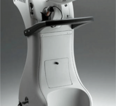
Joseph Imperato, M.D., who practices radiation oncology and radiology in Lake Forest, Ill., uses the Clarity Breast System, an ultrasound from Resonant Medical designed for targeting treatments. According to Dr. Imperato, “Physicians have traditionally relied on the lumpectomy scar to deliver radiation to the tumor cavity, but several studies have found that locating the lumpectomy cavity based solely on this scar may result in a partial miss in over 50 percent of the cases. The lumpectomy cavity may change shape or move in the days and weeks following surgery, and the inconsistent setup of patients on the radiation table may also contribute to this problem. A practical solution is to visualize the cavity on a daily basis using image-guided radiation therapy.”
Due to the issue of variations in the size and shape of the lumpectomy cavity, imaging must take place at both the treatment planning stage and on occasion prior to each consecutive radiation treatment to accurately target the tumor bed, noted Dr. Imperato.
“Currently, CT is used during treatment planning to image the target, but it is limited in its ability to distinguish the lumpectomy cavity from normal tissue,” he said. As a result, “radiation therapists tend to increase the treatment margins to make sure they are encompassing their target.”
There is currently no standard in breast irradiation for daily monitoring of tumor cavity motion. The use of daily CT imaging is not appropriate due to the daily exposure to extra radiation dose.
The Clarity Breast System addresses the issue of lumpectomy cavity movement at both the planning and treatment phases.
“With Clarity, the radiation therapist compares an ultrasound-CT fusion taken at the time of treatment planning with the daily ultrasounds to get an actual visual image and location of the tumor cavity. Once a fused image of ultrasound and CT is achieved, the therapists can then conduct daily imaging with ultrasound-only to assure they are properly delivering radiation to the target that is pre-established with ultrasound/CT fusion. Imaging the tumor cavity every day allows the therapist to adjust the radiation depending on the location of the target,” said Dr. Imperato. “This process ensures greater precision in treatment, which is extremely important in partial breast irradiation, with no additional radiation dose.”
He added, “When it is used properly, the image from the Clarity system allows the radiation oncologists see both the bony and soft tissue anatomy. The image allows the physicians to tighten treatment margins because they are delivering radiation based on a ‘real time image of the tumor cavity.”
“For patients receiving partial breast irradiation, it is very important that the radiation be delivered directly to the lumpectomy cavity to ensure they are getting the entire target,” said Dr. Imperato. “It is important for radiation therapists to have the greatest possible precision when delivering radiation to the target that includes both the soft-tissue image of an ultrasound and a CT image.”
With the assistance of imaging to track the position of lumpectomy cavity, physicians can deliver radiation faster and more accurately. While additional clinical trials are needed to test the safety and effectiveness of new techniques such as APBI, these new techniques hold the promise that partial breast irradiation may increase quality of life for many women with breast cancer.
1. “Partial breast irradiation or whole breast radiotherapy for early breast cancer: A meta-analysis of randomized controlled trials.” A. Valachis, D., Mauri, N. P. Polyzos, D. Mavroudis, V. Georgoulias, G. Casazza; University of Crete, Heraklion, Greece; PACMeR: Oncology and Obstetrics and Gynaecology, Lamia, Greece; University of Milan, Milan, Italy J Clin Oncol 27:18s, 2009 (suppl; abstr CRA532). 2009 ASCO Annual Meeting.
2. “Partial-Breast Irradiation Therapy With MammoSite Appears to Offer Similar Results as Whole-Breast Irradiation Therapy.” F. Vicin, et al, published in the American Society of Clinical Oncology Annual Meeting, June 2006, Abstract 529.
3. APBI Trial. Stanford University Medical Center. http://www.apbitrial.org

 October 01, 2023
