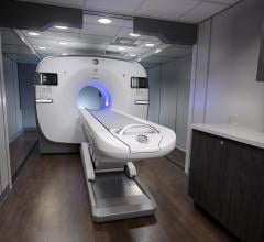
This image, acquired on Siemens' Biograph PET/CT, shows intracerebral metastases.
Could the treatment of Alzheimer’s disease one day become as routine as treating high cholesterol? Instead of a simple blood test to determine whether a patient should take Lipitor to lower his cholesterol, an early diagnostic-PET scan would tell you if you need treatment to prevent the onset of Alzheimer’s. These are the types of treatments that at one point in time may have seemed far-fetched, but with advances in nuclear medicine they are fast becoming a reality.
Advances in hybrid imaging tools involving positron emission tomography (PET) have enabled visualization of chemical markers for disease, which in turn results in better diagnosis, differential diagnosis and discovering disease at earlier stages. The end result may even mean the development of drugs to prevent disease altogether if one shows a pattern of predisposition at a molecular level.
“Nothing compares to PET and single photon emission computed tomography (SPECT) in terms of being able to detect the smallest concentrations of a material within the body.” said Sandi Kwee, M.D., assistant professor at the University of Hawaii and nuclear medicine physician at Hamamatsu/Queen’s PET Imaging Center, The Queen’s Medical Center, in Honolulu. “Radioactive tracers can be detected at sub-nanomolar concentrations within the body, while current magnetic resonance-based methods of tracer detection require much higher concentrations. Furthermore, several engineering obstacles must be overcome to allow PET instrumentation to function properly in the presence of a high-magnetic field gradient.”
Imaging agent vs. imager
In nuclear medicine, visualizing radioactive and chemical activity greatly depends on the specificity imparted by the chemical agent or tracer. “[The early detection of disease] certainly is about the specificity of the equipment, but it is really more about the specificity of the agent,” explained Gene Saragnese, vice president of molecular imaging and computed tomography (CT) for GE Healthcare.
Still, the imaging tools must deliver exceptional image quality, fast processing speeds and accurate data in order to interpret molecular interaction. To this end, Saragnese touted GE’s most advanced hybrid-imaging instrument. “Discovery VCT is the highest specificity in the industry and most advanced CT in the industry,” said Saragnese. The system combines the functional capabilities of PET with the speed and resolution of volume CT. “That combination gives you the platform and will let you look at those things earlier than any other system in the industry. In the area of the PET system, it really is about specificity. It is about acquiring information from the body very rapidly and very accurately. It has the highest resolution, lowest dose we have out there today, and it has the capabilities to do advanced applications beyond those of other CTs.”
New tracers light up
Whether early diagnostics relies more on imaging equipment or on biomarkers for diagnostics is yet unclear. But what is certain is that nuclear medicine is driving new developments in both the chemical tracers and the equipment that visualize them. The end result equates to significant improvements in healthcare thanks to early detection
of disease. This has been most evident in the fields of neurology
and oncology.
Dr. Kwee has explored a new investigational imaging agent for cancer using Philip’s Gemini TF 64 PET/CT system. The Gemini TF 64 has “TruFlight” PET technology, which is Time-of-Flight reconstruction for short scan times and high image quality. Dr. Kwee explained, “We have had experience with fluorine-18 fluorocholine, which is an investigational tracer at the moment. While the jury is still out for the tracer, it does seem better than fluorodeoxyglucose for detecting some cancers, particularly prostate cancer. One nice thing about this tracer is that it is taken up by tissues faster than fluorodeoxyglucose, which means imaging can be performed right after injection. With fluorodeoxyglucose, patients have to wait at least one hour after tracer injection before the actual PET/CT scan is performed.”
Unraveling the mystery of Alzheimer’s
Intense research has recently focused on discovering better diagnostics and treatments for Alzheimer’s disease, which today affects 5 million Americans, is the seventh-leading cause of death in the U.S. and will proliferate as the nation’s population ages.
In a study at Stanford University under the direction of Christopher Contag, Ph.D., associate professor of pediatrics, microbiology and immunology, researchers found a short protein that sticks to colon cells in the early stages of cancer. Using a miniature microscope called the Cellvizio GI, which was developed by Mauna Kea Technologies, the researchers were able to visualize a fluorescent patch created by short proteins. These proteins attach to colon cells in the early stages of cancer after being sprayed into the colon via a fluorescent beam. Initial studies detected 82 percent of the polyps that were considered cancerous. Looking at additional proteins in conjunction with the one discovered, Contag believes that this technique will become highly accurate.
Similar leaps in early detection and differential diagnosis have been made in the field of neurology. Researchers involved in a large multi-institutional study1 used PET imaging with fluorodeoxyglucose (FDG) and were able to classify different types of dementia by measuring the cerebral metabolic rate of glucose (CMRglc) in various areas of the brain. The area of the brain where a decrease in metabolic rate occurred indicated different types of dementia. Those examined included Alzheimer’s disease, frontotemporal dementia and dementia with Lewy bodies, and, more than 94 percent of the time, the disease type was correctly classified using this technique.
Gary Small, M.D., professor of psychiatry and aging at UCLA Femel Institute, has also contributed to new developments in the early detection of Alzheimer’s.2 He employed Siemen’s ECAT HR and EXACT HR tomography, reportedly one of the most widely used PET systems in the world, in his research on Alzheimer’s. Based on the past discovery that certain abnormal protein deposits called amyloid senile plaques and tau neurofibrillary tangles are hallmarks of Alzheimer’s disease, he used a new tracer, FDDNP, (2-(1-6-[(2-[F-18] fluoroethyl)(methyl)amino]-2-naphthylethylidene) malonitrile, to visualize the pattern of radioactivity within the brain using FDDNP-PET scanning.
“What has been predicted and what is remarkable in terms of this strategy has been people with mild cognitive impairment won’t get Alzheimer’s disease for years because we can see a pattern of deposition,” said Dr. Small. “And in our initial follow-up studies, we find that the disease gets worse as people get more forgetful. We see more of the protein marker on the PET scan. Those are the kind of characteristics that you would watch for in a surrogate marker or biological marker that would tag the disease. The next step will be to try a treatment to see if we can slow down the progression of the disease and also have an impact on the marker.”
Interestingly, a new report3 released in late April of this year highlighted the findings of an investigational pill, tarenflurbil (Flurizan) that led to slower declines in global function and in the ability to engage in normal daily activities compared with placebo. However, beginning use of the drug in the early stages of Alzheimer’s disease was crucial to its effectiveness in slowing down the progression of the disease. Tarenflurbil inhibits the gamma-secretase enzyme that produces beta-amyloid protein, thereby blocking the production of the AB42 species of beta-amyloid that forms fibrillary plaques in the brains of Alzheimer’s patients. As a result, the drug prevents deposits of amyloid plaques, but does not appear to destroy existing plaques.
The developments in preventive medication, better diagnosis and early detection also mean improvements in the cost of healthcare. Saragnese noted, “When you look at the healthcare industry today, we have demographics that show that we have more people who are aging and living longer. And the cost of healthcare continues to escalate. And it’s really important that manufacturers and providers of care look at the efficacy of healthcare. So, molecular diagnostics, or genomics and proteomics, I think will allow the identification and categorization of patients in such a way that we can identify high-risk patients and watch them more closely.”
Through nuclear medicine, researchers are finding that diagnosis and treatment of dementia, cancer and other diseases is possible at an earlier, more treatable stage and could one day become as manageable as treating high cholesterol. <
References
1. Tsui WH, Herholz K, Pupi A, et al. Multicenter standardized 18F-FDG PET diagnosis of mild cognitive impairment, Alzheimer’s disease, and other dementias. Journal of Nuclear Medicine March 2008.
2. Small G, Kepe V, Ercoli L, et al. PET of brain amyloid and tau in mild cognitive impairment. 2006; 355:2652-2663. I couldn't find the journal for this one.
3. Black S, et al. Efficacy and safety of tarenflurbil, a selective amyloid beta42-lowering agent, in Alzheimer’s disease (AD): A phase 2 trial of up to 24 months of treatment. Neurology 2008; 70:A392-A393.


 November 18, 2025


