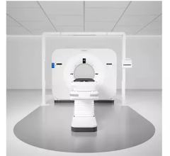
University of Wisconsin Carbone Cancer Center Becomes First in the World to Treat Patients With MRIdians Soft-Tissue-Tracking-Beam Control
September 12, 2014 — The MRIdian system from ViewRay is now being used to treat patients at the University of Wisconsin Carbone Cancer Center in Madison, Wis., the second clinical group in the world to treat patients with magnetic resonance imaging (MRI)-guided radiation therapy. With the treatment, the center also became the first clinic in the world to employ the soft-tissue tracking beam control feature of ViewRay’s MRIdian system.
The MRIdian system is the world’s first and only MRI-guided radiation therapy system. It provides continuous imaging during treatment so clinicians are able to see where the radiation dose is being delivered and adapt to changes and movement in the patient’s anatomy in real-time. The system tracks soft-tissues of the tumor directly, in fast planar images, then automatically compares the target to the plan and only allows treatment when the target is in range.
“We had a patient with very limited treatment options,” said Michael Bassetti, M.D., Ph.D., assistant professor of radiation oncology at the University of Wisconsin Carbone Cancer Center. “The size and location of this patient’s liver tumor combined with the large observable motion prevented the safe delivery of treatment with X-Ray based linac systems. With ViewRay’s MRIdian system, we were able to ensure the accuracy of the delivery by directly tracking tumor and normal soft-tissue movement during treatment.”
The first MRI-guided radiation therapy patient at Carbone Cancer Center was a liver cancer patient who was treated with the soft-tissue tracking beam control feature employing a 5 mm margin on a breath-hold scan acquired on the system.
“I am grateful UW-Madison can now provide this remarkable treatment approach to our patients,” said John Bayouth, Ph.D., professor and chief of physics at the University of Wisconsin Carbone Cancer Center. “MRIdian enabled us to meet demanding prescription goals through its ability to track the actual tumor and surrounding normal tissues in real-time. We minimized the radiation dose to the bowel and normal liver while verifying the dose delivered to the tumor during treatment delivery. No longer do we have to assume what is happening inside the patient during treatment, now we are able to see changes and respond accordingly.”
“We are excited to be one of the first centers to use MRI-guided radiation therapy in clinical practice,” said Paul Harari, M.D., professor and chairman of the Ddpartment of human oncology at the University of Wisconsin School of Medicine and Public Health. “Continual MR image-guidance and control is unique to the MRIdian system with the potential to change the way clinicians treat cancer patients.”
For more information: www.viewray.com


 November 04, 2025


