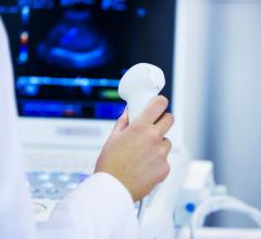
April 6, 2011 – Ultrasound is often used as a first-line diagnostic exam to quickly and safely diagnose a range of patient conditions. At this year’s American College of Cardiology (ACC) annual meeting in New Orleans Toshiba America Medical Systems highlighted new enhancements to its cardiac and shared service ultrasound systems that are designed to enhance cardiac ultrasound imaging.
For its flagship cardiac system, Aplio Artida, 3-D wall motion tracking and tissue enhancement technologies are now available. 3-D wall motion tracking offers a new era of dyssynchrony imaging and advanced regional wall motion assessment. It aids electrophysiologists in optimizing pacemaker placement and function. It also shows 3-D ejection fraction, volumes and regional and global strain function. A Toshiba-exclusive software, tissue enhancement has the ability to improve image uniformity and endocardial border delineation, especially in difficult-to-scan patients.
Available on Toshiba’s shared service ultrasound systems, Aplio MX, Aplio XG and Xario XG, the new Auto IMT feature calculates the intima-media thickness of the carotid artery, helping clinicians determine a patient’s risk for cardiovascular disease. It can determine the thickness of the near and far arterial walls from three segments of the carotid artery: at an optimal angle of incidence and two complementary planes. The software uses the collected images following the American Society of Echocardiography (ASE) consensus statement for diagnosis of cardiac risk in certain asymptomatic populations.
Also, the company showed the new adult motor-driven transesophageal (TEE) probe, which improves the diagnosis of numerous cardiac conditions in difficult-to-scan patients.
For more information: www.medical.toshiba.com

 July 19, 2024
