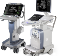
December 19, 2007 - Toshiba Medical Systems Europe recently announced the company’s newest product, the Artida ultrasound system, designed to meet the demands of the growing cardiac 4D market.
With Artida’s real-time multi-planar reformatting capabilities, physicians can reportedly quantify global and regional LV function, including LV ejection fraction, volume and severity of regurgitation. Arbitrary views of the heart not available in 2D imaging are also obtained that can help with surgical planning, according to the company.
The Artida was shown for the first time at the ESC Congress in Vienna, Austria.
The Artida has the ability to track and display myocardial motion in 3D images. This Wall Motion Tracking feature allows the user to obtain angle-independent, quantitative and regional information about myocardial contraction. This ability to identify wall motion defects will greatly improve Cardiac Resynchronization Therapy (CRT) using pace makers by determining who will be a responder to CRT and who will probably not. It will also help physicians optimize the pace maker setting.
The system also features a SmartCore engine, which reportedly employs the distributed processing power of more than 80 processor cores interconnected by a fast digital system interface, a MultiCast Beamformer, which uses advanced digital signal processing to reportedly control the shape of the ultrasound beam more precisely and flexibly than in comparable systems and SmartSlice functions that allow physicians to cut, slice and position the 4D volume quickly and conveniently, according to Toshiba.
For more information: www.medical.toshiba.com


 March 19, 2025
