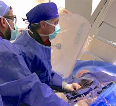
April 1, 2008 - Toshiba America Medical Systems Inc. introduced its Advanced Image Processing (AIP) technology for its Infinix X-ray product line designed for improved device and vessel visualization for interventional procedures at this year's American College of Cardiology (ACC) annual meeting in Chicago.
"AIP allows us to image bariatric patients, which was difficult to do before. It also gives us reduced background noise and improved device and vessel visualization, which allows us to increase workflow and provide better patient care during our interventional procedures," said John S. Wilson, M.D., medical director, cardiovascular services, Washington Hospital.
According to the manufacturer, benefits of the technology are engineered to include:
- Programmable image display: The system works with new software to allow clinicians the ability to optimize image viewing based on clinical preferences.
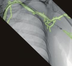
- Reduced background noise for fluoroscopic and digital acquisition imaging: This new processing is designed to compress background noise and enable physicians to better see vasculature and cardiac devices.
- Improved device and vessel visualization: AIP is supposed to enhance contrast, improving the ability to see vessels and devices without increasing the overall image noise.
- Improved resolution in dark areas: AIP combined with high-resolution flat panel detectors allow vessels near dense anatomical areas may be seen more easily.
- Improved dynamic range: This technology is said to reduce the blurring effect that can sometimes occur in areas of high contrast (i.e., heart tissue to lung field), providing a much clearer image for physicians to review.
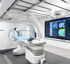
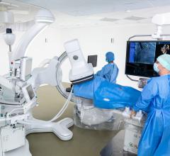
Toshiba showcased AIP in conjunction with its Infinix VF-i/SP X-ray system, a universal cardiovascular system designed to accommodate diagnostic and interventional procedures.
Developed based on the Infinix-i series platform, the new floor-mounted, single plane system features a multi-axis positioner with extremely versatile movement for unprecedented patient access and anatomical coverage, including head-to-toe, finger tip-to-finger tip coverage. These features aim to allow clinicians to complete procedures more quickly and comfortably than traditional systems, reducing procedure times and improving overall departmental workflow. In addition, a high-resolution 12x16 inch flat panel detector is said to provide the uniform, distortion-free images required for the full range of cardiovascular studies.
For more information: www.medical.toshiba.com.


 May 17, 2024
