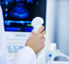
November 29, 2007 - According to a study presented at the RSNA, ultrasound-guided nonsurgical therapy can significantly reduce pain from calcific tendonitis of the rotator cuff and restores mobility.
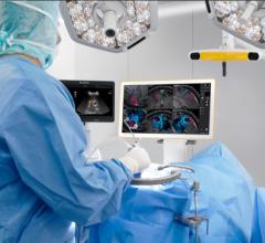
Ultrasound-guided percutaneous therapy is a 10-minute procedure in which the shoulder is anesthetized and, with ultrasound guidance, a radiologist injects a saline solution into the rotator cuff to wash the area and break up the calcium. A second needle is used to withdraw the calcium residue. Recovery time is about an hour and calcifications that are completely treated do not return.
"This is a quick, successful and inexpensive therapy for tendon calcifications," said Luca M. Sconfienza, M.D., from the Department of Radiology at A.O. Ospedale Santa Corona in Pietra Ligure and the Department of Experimental Medicine at the University of Genova in Italy. "It provides significant and long-lasting reduction of symptoms."
"Calcifications can break up on their own. Unfortunately, this can take from a few months to several years," Dr. Sconfienza said. "This means the pain could persist for years before its spontaneous resolution."
When left to break up on their own, untreated calcifications can spread calcium along the tendon and lodges in the subacromial bursa, a fluid sac that helps lubricate the tendon.
The study involved 1,607 women and 938 men (mean age 42) with calcific tendonitis. All of the patients had shoulder pain that was unresponsive to previous medical treatment. One-year follow-up was reported for 2,018 of the patients in the study.
The results showed that in 71.7 percent of the patients, the calcification was fully aspirated in one treatment with a considerable reduction in pain and significant improvement to mobility of the affected limb. In 23.6 percent of patients, a second procedure was performed because of the presence of more than one calcification. In 3.8 percent of patients, the calcification had dissolved or moved before treatment could take place. In 0.9 percent of patients, no resolution of symptoms occurred because of the presence of a tendon tear.
Dr. Sconfienza says that theoretically the procedure could be performed in any hospital or clinic that has ultrasound equipment with a superficial probe.
For more information: www.rsna.org


 March 19, 2025
