Image courtesy of MR Solutions.
May 23, 2019 — Researchers from Bourgogne University in Dijon, France, showed that use of superparamagnetic iron oxide nanoparticles (NPs) using multimodal positron emission tomography/magnetic resonance imaging (PET/MRI) offers promising improvements in nuclear imaging capabilities. The paper is published in the American Chemical Society journal ACS Omega.1
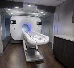
Using an MR Solutions cryogen-free, 3 Tesla (3T) scanner with a PET clip on the sequential images were found to be highly beneficial providing high spatial resolution, high sensitivity and high technical maturity with a low radiation dose.
Prof. Nadine Millot, principal investigator on the study commented, “The combination of simultaneous MRI/PET imaging is very attractive because it allies the high resolution of MRI with the high sensitivity of PET to obtain accurate anatomical and quantitative information at the same time.” This pioneering imaging agent highlights activity in the liver, spleen, lungs and kidneys before being gradually excreted from the body, with no cytotoxicity observed.
This is the first successful grafting of PEG and MANOTA chelator on prefunctionalized iron oxide NPs synthesized under continuous hydrothermal conditions for bimodal PET/MRI imaging and shows the potential benefits.
For more information: www.pubs.acs.org
Reference
1. Thomas G., Boudon J., Maurizi L., et al. Innovative Magnetic Nanoparticles for PET/MRI Bimodal Imaging. ACS Omega, published online Feb. 5, 2019. https://doi.org/10.1021/acsomega.8b03283


 July 31, 2024


