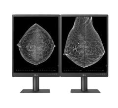
72-year-old woman recalled from screening mammography for architectural distortion in left breast. A. Craniocaudal and B. mediolateral oblique digital breast tomosynthesis images demonstrate two areas of AD in upper outer left breast (circles) that persisted on additional diagnostic tomosynthesis images (not shown). C. Transverse greyscale ultrasound image of upper outer breast shows irregular hypoechoic mass with associated AD at 12:30 (arrow), corresponding to posterior AD. No ultrasound correlate was identified for anterior AD. Ultrasound-guided biopsy of posterior AD revealed malignancy (invasive lobular carcinoma). Tomosynthesis-guided biopsy of anterior AD yielded benign pathology (stromal fibrosis).
July 28, 2022 — According to ARRS’ American Journal of Roentgenology (AJR), for patients with multiple architectural distortion (AD) identified on digital breast tomosynthesis (DBT), biopsy of all areas may be warranted, given the variation of pathologic diagnoses across AD in individual patients.
“Multiple AD, compared with single AD, was significantly more likely to yield high-risk pathology, but there was no significant difference in yield of malignancy,” wrote corresponding author Lilian C. Wang, MD, of Northwestern Medicine. “In patients with multiple AD, multiple ipsilateral or contralateral AD commonly varied in pathologic classification: benign, high risk, or malignant.”
Wang and colleagues’ retrospective study included 402 patients (mean age, 56 years) who underwent image-guided core needle biopsy of AD visualized on DBT between April 7, 2017, and April 16, 2019. Patients were grouped in the single or multiple AD cohort based on the presence of distinct AD areas noted in the clinical report, while the pathologic diagnosis for each AD was based on the most aggressive pathology identified on either biopsy or surgical excision (if performed).
Ultimately, compared with single AD, multiple AD showed a higher frequency of high-risk pathology (53.0% vs. 32.5%, p=.002) but no significantly different frequency of malignancy on a per-lesion or per-patient basis (31.8% vs. 28.2%, p=.56). In 8/24 patients with ≥2 ipsilateral biopsied AD, the ipsilateral areas varied in terms of most aggressive pathology; in 5/10 patients with contralateral biopsied AD, the contralateral areas varied in most aggressive pathology.
“To our knowledge,” the authors of this AJR article contended, “this is the first study to compare pathologic outcomes between patients with single and multiple AD visualized by DBT.”
For more information: www.arrs.org
Related Digital Breast Tomosynthesis Content:
Today's Mammography Advancements
Digital Breast Tomosynthesis Spot Compression Clarifies Ambiguous Findings
AI DBT Impact on Mammography Post-breast Therapy
ImageCare Centers Unveils PINK Better Mammo Service Featuring Profound AI
Radiologist Fatigue, Experience Affect Breast Imaging Call Backs
Fewer Breast Cancer Cases Between Screening Rounds with 3-D Mammography


 July 29, 2024


