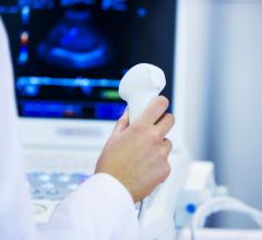
April 7, 2011 – At the recent American College of Cardiology (ACC) Annual Scientific Session in New Orleans, Siemens showcased several ultrasound products. Among the systems highlighted were the 1.6 release of its Acuson SC2000 volume imaging ultrasound system and IN Focus Technology, derived from the Acuson Sequoia ultrasound system.
The Acuson SC2000 system is the only system that delivers the same detail resolution across the entire image without sacrificing frame rates. The IN Focus Technology acquires and processes information at an unprecedented 2.88 gigabytes per second, faster than any echocardiography system in the world. This leads to never-before-seen detail and contrast resolution throughout the entire field of view delivering more clinically relevant information, ultimately benefiting a patient’s diagnosis.
An advancement to the Acuson Sequoia coherent image formation technology, IN Focus Technology enables the user to focus on the entire field of view instead of a single focal zone revealing more detailed information in one image. By using the power of 64 parallel receive beams, it dramatically improves image quality at all depths without any user intervention. This helps display the cardiac structure, motion and blood flow information for superior and efficient diagnostic imaging.
The Acuson SC2000 system exclusively features the eSie Measure Workflow Acceleration package. This application provides fully automated measurements for routine echo exams, which increases workflow efficiency, as well as the reproducibility and quality of each exam, while additionally addressing the issue of repetitive stress injuries in sonographers. Also, eSieScan was integrated into the system to further streamline exam workflows on both the user level and in the entire lab. These protocols bring higher reproducibility and quality standards to the echocardiography workflow by increasing the consistency of results and ensuring that exams are complete.
The feature is customizable according to user or department requirements, and dramatically reduces the need for user interaction and the number of keystrokes during the imaging process.
“One of the most significant benefits using eSieScan workflow protocols is the reduction of exam times,” said Adrien Ntinunu, Sonographer at the Ohio State University, Columbus, Ohio. “Typically, manual measurements can take an average of 20-25 minutes. The eSie Measure Workflow Acceleration package has saved us significant time in each exam. It is useful for every patient, especially for aortic insufficiency and aortic stenosis patients.”
Called “Echo in a Heartbeat,” the company’s real-time full-volume imaging capabilities on the Acuson SC2000 system allow wider patient access by delivering vastly more diagnostic information. In one heart cycle and without stitching or ECG gating, it acquires the full volume of the heart at a 90 by 90 degree angle and 16-centimeter depth at up to 40 volumes per second. This includes volumetric color flow and accurate volume quantification for the left and right ventricle. It is easily integrated into a routine adult echocardiography exam, and offers new workflow pathways to improve diagnostic confidence and efficiency.
In order to take advantage of 4-D imaging, a number of technologies have been integrated into the real-time full-volume ultrasound system. After acquiring a volume, eSie LVA LVA (Left Ventricle Analysis) Volume automatically draws the contour of the left ventricle from the volume dataset, generating ejection fraction (EF) and volume data in as little as 15 seconds. The system compares the clinical case on hand with results from a database containing thousands of clinical cases.
This gives physicians a better overview of available patient data, eSie LVA supports the standard American Heart Association (AHA) segmentation for 16 and 17 segments, standardizing exam protocols between computed tomography (CT), magnetic resonance imaging (MRI) and molecular imaging. Ultimately, this offers physicians more comprehensive information about their patients.
The company also showcased knowledge-based workflow applications on various platforms, which are designed to automate measurements for rapid, accurate and reproducible results.
• syngo Velocity Vector Imaging™ (VVI) technology uses individual vectors to display direction and relative velocity of tissue from frame to frame to instantaneously measure motion at any point in the cardiac cycle. This allows easy information gathering n for a variety of applications, including rapid assessment of ventricular synergy in heart failure.
• syngo Auto Left Heart (Auto LH) technology automatically generates left atrial and left ventricular volumes and ejection fractions rapidly and reliably.
• Rapid Stress volume stress echo application features full-volume acquisition in one single heartbeat per stage and auto-extraction of reference planes for comparison. The resulting volume stress workflow enables the only volume stress echo solution for patients with arrhythmia.
For more information: www.siemens.com/healthcare

 July 19, 2024
