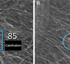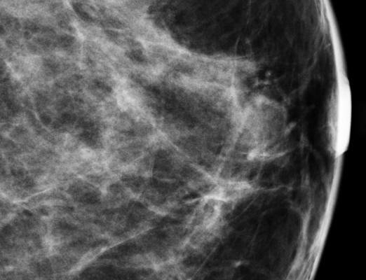
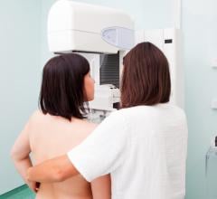
December 13, 2012 — Researchers reviewing the records of approximately 250,000 women enrolled in an integrated healthcare delivery system found that increased CT (computed tomography) utilization between 2000 and 2010 could result in an increase in the risk of breast cancer for certain women, including younger patients and those who received repeat exams. According to the study, which was presented at the 98th annual meeting of the Radiological Society of North America (RSNA), nuclear medicine examinations may also contribute to increased breast cancer risk.
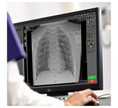
CT uses ionizing radiation in the form of X-rays to produce cross-sectional images of the body. In nuclear medicine imaging, a radiopharmaceutical is delivered inside the body to help visualize internal organs.
"When a woman undergoes CT or nuclear medicine imaging of her chest, abdomen or spine, her breast tissue will absorb some radiation," said senior author Rebecca Smith-Bindman, M.D., professor of radiology and biomedical imaging at the University of California, San Francisco. "Breast tissue is one of the tissues in the body known to be sensitive to developing cancer as a result of radiation exposure."
The study, led by Ginger Merry, M.D., M.P.H., breast imaging fellow at Prentice Women's Hospital – Northwestern Memorial Hospital in Chicago, found that among the system's female enrollees, CT utilization increased from 99.8 CT scans per 1,000 women in 2000 to 192.4 CT scans per 1,000 women in 2010 (an annual increase of 6.8 percent). In 2010, 46 percent of those CT examinations exposed the breast to radiation. Nuclear medicine imaging decreased from 39.3 scans per 1,000 women in 2000 to 27.5 scans per 1,000 women in 2010 (a 3.5 percent annual decline); however, in 2010, 84 percent of nuclear medicine studies exposed the breast to radiation.
"Until now, the impact of this increased use of imaging on radiation exposure to breast tissue and the subsequent risk of breast cancer has not been known," Smith-Bindman said. "Our goal was to quantify imaging utilization and radiation exposure to the breast among women enrolled in an integrated healthcare delivery system, and to use these data to determine the imaging-related risk of breast cancer from those studies."
The research team collected CT dose information from 1,656 patients who underwent CT examinations that exposed the breast to radiation and, using a new automated computational method, estimated the patients' effective radiation dose and the amount of radiation absorbed by the breast. The team also analyzed the radiopharmaceutical volume and associated radiation exposure used in 5,507 nuclear medicine exams that exposed the breast to radiation.
"We found that the estimated breast radiation doses from CT were highly variable across patients, with the highest doses coming from multiple-phase cardiac and chest CT examinations, where successive images of the organ being studied are captured," Smith-Bindman said.
The researchers then estimated the women's imaging-related risk of breast cancer and compared it to their underlying risk of developing breast cancer. Each woman's 10-year imaging-related risk of developing breast cancer, beginning 10 years after her exposure to imaging and based on her age at exposure, was estimated using the breast-specific radiation data and a statistical risk model. A women's underlying risk of developing breast cancer was estimated based on data collected by the National Cancer Institute-funded Breast Cancer Surveillance Consortium.
"Young women receiving several chest and or cardiac CTs had the greatest increased risk of developing breast cancer at approximately 20 percent," said Diana Miglioretti, Ph.D., study co-author and senior investigator at the Group Health Research Institute. "A 15-year-old girl with no risk factors for breast cancer would double her 10-year risk of developing breast cancer at 25."
To lower imaging-related risk of developing breast cancer, Smith-Bindman said imaging providers should analyze the radiation doses associated with each exam, reduce the use of multiphase protocols and employ dose reduction software wherever possible to minimize exposures.
"If imaging is truly indicated, then the risk of developing cancer is small and should not dissuade women from getting the test they need," she said. "On the other hand, a lot of patients are undergoing repeat chest and cardiac CT, many of which aren't necessary. Women, and particularly young women, should understand there is a small but real potential risk of breast cancer associated with cardiac and chest CT, and the risk increases with the number of scans."
For more information: www.rsna.org
 July 29, 2024