March 4, 2008 - By using a dual-source technique on a single computed tomography (CT) system, a research team at the Medical University of South Carolina’s (MUSC) Heart & Vascular Center was able to make a comprehensive diagnosis of heart disease, according to a report published in the current issue Circulation.
The team, led by Balazs Ruzsics, M.D., Ph.D.; Eric Powers, M.D., medical director of MUSC Heart and Vascular Center; and U. Joseph Schoepf, M.D., director of CT Research and Development, explored how CT scans can now detect blocked arteries and narrowing of the blood vessels in the heart in addition to poor blood flow in the heart muscle.
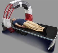
The single-scan technique would also provide considerable cost savings, as well as greater convenience and reduced radiation exposure for patients. For their approach, the MUSC physicians used a Dual-Source CT scanner. The MUSC scanner was the first unit worldwide that was enabled to acquire images of the heart with the ‘dual-energy’ technique. While the CT scan "dissects" the heart into thin layers, enabling doctors to detect diseased vessels and valves, it could not detect blood flow. The MUSC researchers added two X-ray spectrums, each emitting varying degrees of energy like a series of X-rays, to gain a static image of the coronary arteries and the heart muscle. This dual-energy technique of the CT scan enables mapping the blood distribution within the heart muscle and pinpointing areas with decreased blood supply.
All this is accomplished with a single CT scan within one short breath-hold of approximately 15 seconds or less. In addition to diagnosing the heart, the CT scan also permits doctors to check for other diseases that may be lurking in the lungs or chest wall.
MUSC, like most cardiovascular centers, had traditionally relied on several imaging modalities, such as cardiac catheterization, nuclear medicine or magnetic resonance (MR) scanners.
"This technique could be the long coveted ‘one-stop-shop’ test that allows us to look at the heart vessels, heart function and heart blood flow with a single CT scan and within a single breath-hold," said Dr. Schoepf, the lead investigator of the study.
Based on their initial observations, Heart & Vascular Center physicians have launched an intensive research project aimed at systemically comparing the new scanning technique to conventional methods for detecting decreased blood supply in the heart muscle.
For more information: www.musc.edu or www.muschealth.com


 August 09, 2024


