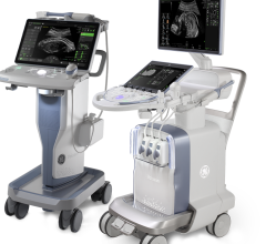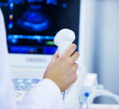
February 28, 2017 — Philips announced 510(k) clearance from the U.S. Food and Drug Administration (FDA) to market its ElastQ Imaging capability, further expanding the functionalities of its Epiq family of ultrasound systems.
ElastQ Imaging enables simultaneous imaging of tissue and assessment of its stiffness, which is essential for the diagnosis of various liver conditions. With ElastQ Imaging, clinicians have a comprehensive solution to assess and diagnose liver conditions without the pain or expense of a liver biopsy. Using shear wave elastography to focus sound waves to assess soft tissue stiffness, ElastQ Imaging is non-invasive, reproducible and easily executed.
Liver disease, which includes hepatitis B and C, liver cancer and cirrhosis, is a growing global health issue due in part to rising obesity rates and an aging population. Non-alcoholic fatty liver disease affects approximately 20 percent of the global population [1]. According to the World Health Organization (WHO), total deaths worldwide from cirrhosis and liver cancer rose by 50 million per year over the last two decades, and many cases continue to go undetected [2].
To determine the stage of liver disease and damage, a liver biopsy is typically performed by extracting a small piece of liver tissue for microscopic examination. Research suggests that instead of costly and painful biopsy procedures, ultrasound exams using shear wave elastography could become routine for assessing liver disease status and may reduce or avoid the need for conventional liver biopsies [3].
Philips ElastQ Imaging shear wave elastography offers:
- Larger field of view or region of interest (ROI) than competitors, according to Philips;
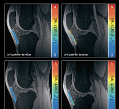
- Color-coded quantitative assessment of tissue stiffness;
- Real-time feedback and intelligent analysis; and
- Quantitative measurements with multiple sample points.
In addition to ElastQ Imaging, Philips' ultrasound liver solution is comprised of a suite of features that make it a powerful tool for clinicians, including:
- PureWave transducer technology, delivering superb image quality and increased penetration in technically difficult-to-image patients;
- Contrast enhanced ultrasound, providing confidence in liver lesion detection and characterization; and
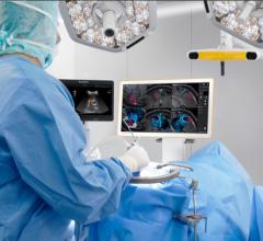
- Image fusion and navigation with anatomical intelligence, increasing the power of real-time ultrasound exams through multimodality fusion and interventional guidance.
Philips’ ultrasound liver solution combines a routine ultrasound imaging exam of the liver anatomy with targeted tissue stiffness values, allowing clinicians to obtain a baseline assessment without the wait time associated with tissue biopsy. For patients, the non-invasive ultrasound exam is more comfortable than a traditional liver biopsy that requires the use of needles. The exam results from ultrasound are also available instantly, which can help reduce apprehension patients may feel in waiting for test results.
"There is a significant population at risk for liver disease that may not even know it," said Richard G. Barr, M.D., Ph.D., radiologist, Southwoods Imaging, in Youngstown, Ohio. "As a radiologist, I see every day how important liver assessment is becoming, and I'm hopeful that this solution will help patients get the diagnosis, monitoring and treatment they need."
For more information: www.philips.com
References
[1] Sattar N, et al. Non-alcoholic fatty liver disease. BMJ 2014;349:g4596. 34 Dyson J et al. Non-alcoholic fatty liver disease: a practical approach to treatment. Postgrad Med J. 2015 Feb;91(1072):92-101.
[2] (2013, November 4). Global Burden of Liver Disease Substantial. Medscape. Retrieved from http://www.medscape.com/viewarticle/813788#vp_1
[3] Ferraioli G, et al. Point shear wave elastography method for assessing liver stiffness. World J Gastroenterology. 2014 April 28;20(16):4787-4796.


 March 19, 2025
