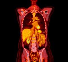
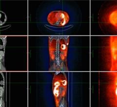
June 18, 2008 - Combined positron emission tomography (PET) and computed tomography (CT) scanning of patients in the early stages of ovarian cancer can enable physicians to determine whether the cancer has spread to nearby lymph nodes without having to perform surgery, according to researchers at the SNM�s 55th Annual Meeting.
As a result, unnecessary surgeries could be reduced, which would also lower morbidity rates and postoperative complications for ovarian cancer patients.
�Our preliminary research indicates that using PET/CT scanning in this way could greatly improve quality of life for many patients with ovarian cancer,� said Luca Guerra, doctor of nuclear medicine, San Gerardo Hospital, Monza, Italy, and lead researcher of the study, 18F-FDG PET/CT Usefulness in Initial Staging of Ovarian Cancer. �PET/CT scans could allow many women to forego major abdominal surgery to determine whether their cancer has spread. It�s a much safer alternative for determining the stages of ovarian cancer.�
More than 20,000 U.S. women will be diagnosed with ovarian cancer this year, according to recent estimates, and more than 15,000 women will die from it. Unlike many other types of cancer, there is currently no reliable screening test to determine whether a woman has ovarian cancer. Physicians usually perform surgery to diagnose and stage ovarian cancer. Although CT and MRI technologies are useful in determining surgery in advanced cases of the disease, both have limited accuracy in determining stages of ovarian cancer.
While systematic lymphadenectomy�surgically removing all of the lymph nodes for testing rather than sampling a small number of the lymph nodes�is more accurate in determining whether the cancer has spread, the surgery takes longer, often requires blood transfusions and can result in life-threatening complications. If all early-stage ovarian cancer patients underwent lymphadenectomy, approximately 75 percent of the surgeries would prove unnecessary.
In their research, Guerra and his team examined results of 30 women diagnosed with ovarian cancer who underwent PET/CT scanning before surgery to determine the stage of their disease. The results indicated that PET/CT staging was correct in 67 percent of patients and more than 98 percent accurate in scanning the lymph nodes of stage I and stage II ovarian cancer patients. These very promising results need to be confirmed in a larger patient population.
While most previous studies that examined the potential of PET technologies have not indicated a definite role in the staging of ovarian cancer, many of the studies involved very small groups of women, and the scanners used were simple PET scanners rather than state-of-the-art, combined PET/CT scanners.
Scientific Paper 440: L. Guerra, M. Arosio, M. Musarra, Department of Nuclear Medicine; with A Garbi, C. Mangioni, Gynaecological Division, San Gerardo Hospital, Monza, Italy; C. Crivellaro, S. Sironi, School of Medicine, all of University of Milan Bicocca, Monza, Italy; C Messa, IBFM-CNR-Institute for Molecular Bioimaging and Physiology, Milan, Italy; F. Fazio, IRCCS San Raffaele Hospital, Milan, Italy, �18F-FDG PET/CT Usefulness in Initial Staging of Ovarian Cancer,� SNM�s 55th Annual Meeting, June 14�18, 2008.
For more information: www.snm.org


 July 02, 2024
