December 12, 2007 - According to research published in the December issue of the Journal of Nuclear Medicine, PET/CT imaging exhibits significantly higher sensitivity, specificity and accuracy than conventional imaging when it comes to detecting malignant tumors in children, and provides doctors with additional information about cancer in children, possibly sparing young patients from being overtreated.
The study found that PET/CT can detect small lymph node lesions diagnosed as negative with conventional (or anatomical) imaging and deny the presence of active disease in soft-tissue masses post-treatment, especially in children with a wide range of malignant cancers.
Researchers reviewed 151 FDG PET/CT exams that were performed on 55 children with noncentral nervous system malignancies (30 patients had lymphoma, cancer that affects the body's lymph nodes and other organs that are part of the body's immune and blood-forming systems).
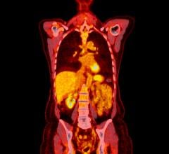
According to the study, PET with CT imaging, with the radiotracer fluorodeoxyglucose (FDG) enables the collection of both biological and anatomical information during a single exam, with PET picking up metabolic signals of body cells and tissues and CT offering a detailed map of internal anatomy. When there were discrepancies between PET/CT and conventional anatomical imaging in analyzing cancer lesions, PET/CT was diagnostically accurate 90 percent of the time. Doctors monitor the radiation dose given to children in imaging exams, generally using lower doses than that of adult patients, to get adequate exam results,
The study says PET is a powerful molecular imaging procedure that noninvasively demonstrates the function of organs and other tissues. When PET is used to image cancer, a radiopharmaceutical (such as FDG, which includes both a sugar and a radionuclide) is injected into a patient. Cancer cells metabolize sugar at higher rates than normal cells, and the radiopharmaceutical is drawn in higher amounts to cancerous areas. PET scans show where FDG is by tracking the gamma rays given off by the radionuclide tagging the drug and producing three-dimensional images of their distribution within the body. PET/CT offers precise fusion of metabolic PET images with high-quality anatomical CT images.
For more information: www. snm.org


 October 30, 2025


