May 24, 2010 - The provision of 3-D/4-D imaging functionality as an integral component of a picture archive and communication systems (PACS) solution is a recent development in the market.
As PACS vendors add advanced visualization to the package, attendees at the Society of Imaging Informatics in Medicine (SIIM) 2010 Annual Meeting, June 3 to 5, in Minneapolis.
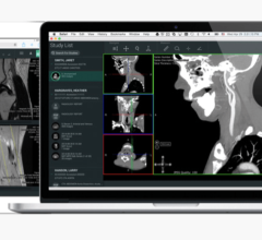
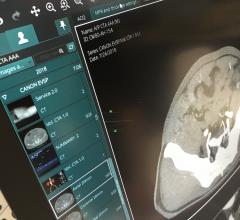
A new image visualization software, Synapse Maximum Intensity Projection and Multi-planar Reformatting (MIP/MPR) Fusion, developed by PACS vendor FUJIFILM Medical Systems U.S.A., is designed for hospitals, imaging centers and radiology groups. The recently FDA-cleared application is the latest addition to the Synapse line of integrated medical informatics products. Synapse MIP/MPR Fusion will be included as a standard application in all Synapse PACS versions 3.2.1 and higher.
The new software includes the standard functionality expected of a maximum intensity projection (MIP) and multiplanar reformation (MPR) solution, and adds several new radiologist workflow tools.
To enhance efficiency, a tool allowing clinicians to readily drag and drop positron emission tomography (PET) images into computed tomography (CT) images, automatic PET/CT fusion images can be created quickly. Additionally, important standard uptake value (SUV) calculations, measurements and displays are designed to be made almost immediately.
Integrated workflow within Synapse can be utilized from any location where Synapse is available, eliminating the need for specialized workstations. This enables clinicians to toggle between MPR, fusion and the orthogonal slices without having to close patient studies or log into another system. Having all medical information readily available from one source saves time and provides for immediate comparisons on a current PACS reading station configuration.
Synapse MIP/MPR Fusion offers customizable reading panels that allow the users to configure their workspace for their reading preferences and workflow needs. Window leveling and slab thickness level presets, customizable overlay types and various image reading formats can be selected and customized.
For more information: www.fujimed.com

 April 18, 2025

