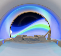
August 22, 2007 - Research published in the academic journal, Chemical Communications, reveals that a new compound may be used in a 'chemically-sensitive MRI scan' to help identify the extent of progression of diseases such as cancer, without the need for intrusive biopsies.
The researchers, who are part of an Engineering and Physical Sciences Research Council (EPSRC) funded group developing new ways of imaging cancer, have created a chemical that contains fluorine. It could, in theory, be given to the patient by injection before an MRI scan. The fluorine responds differently according to the varying acidity in the body, so that tumours may be highlighted and appear in contrast or 'light up' on the resulting scan.
Professor David Parker of Durham University's Department of Chemistry explained, "There is very little fluorine present naturally in the body so the signal from our compound stands out. When it is introduced in this form it acts differently depending on the acidity levels in a certain area, offering the potential to locate and highlight cancerous tissue."
Professor Parker's team is the first to design a version of a compound containing fluorine which enables measurements to be taken quickly enough and to be read at the right 'frequency' to have the potential to be used with existing MRI scanners, whilst being used at sufficiently low doses to be harmless to the patient.
For more information: http://www.dur.ac.uk


 July 25, 2024


