July 29, 2008 - For several years, doctors have known that certain stem cells migrate to the heart after a heart attack, but exactly how they get there and what purpose they serve remained uncertain.
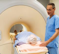
Now for the first time, researchers have tracked the stem cells in mice using magnetic resonance imaging (MRI) from their bone marrow origin to the injured site. This opens up the possibility of finding some therapeutic treatment to direct these cells after a heart attack.
Mesenchymal stem cells, or MSCs, are found in the bone marrow and can differentiate into certain cell types. They have been detected around heart injuries following a myocardial infarction (heart attack), but whether they come to regenerate heart tissue or to promote healing is still under debate.
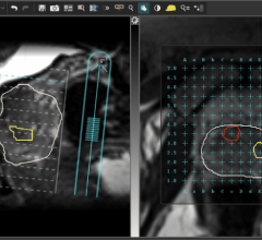
Using a series of MRI scans, Tom Hu and colleagues at the Medical College of Georgia in Augusta, GA, have tracked MS's in a sample of mice. The researchers first transplanted into the bone marrow a few hundred thousand MSCs that had been labeled with both iron-oxide (a molecule that essentially shades out the MRI signal) and a special protein that fluoresces when exposed to blue light. The team then operated on all of the mice, inducing a heart attack in one group. Over the following days, MRI scans showed a gradual darkening around the site of injury in the heart attack group, which was presumably due to the arrival of the labeled MSCs. The researchers validated this migration with fluorescent microscopy.
The goal now is to devise a way to attach an MRI-sensitive marker to MS's in humans who have suffered a heart attack. This would allow doctors to more closely study these cells, and perhaps devise treatments that can control their migration.
For more information: www.aapm.org
Source: American Association of Physicists in Medicine

 April 10, 2025