February 8, 2011 – Researchers at the University of Southern California have developed an algorithm to produce animated magnetic resonance imaging (MRI) scans of body parts or organs in motion. Volume scans of human bodies have a variety of uses in medical diagnosis and research; however, these scans usually do not show the body in movement.
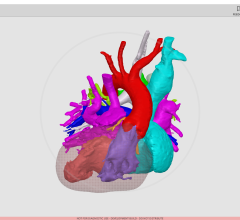
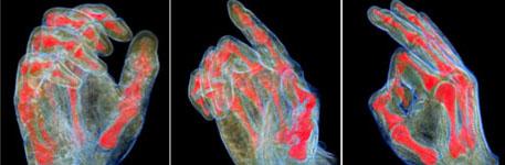
The new algorithm can reconstruct an animated representation of any area of the body. Skeletal joints of the reconstructed representation can be moved with a 3-D animation program and the soft tissues deform interactively and realistically according to the MRI scan data. The preliminary application is for orthopedic evaluation, but the software could also be applied for organ movement, such as the heart or lungs.
The underlying approach is an intelligent example-based interpolation technique called volume blend deformation. The challenge in applying this technique is accurate registration of scans in different poses. This required the development of a new hierarchical skeleton-guided registration step. Although the registration requires many hours of pre-computation, the final result can be animated interactively.
The new algorithm will be particularly valuable in visualizing problems related to movement, since it visualizes the internal structures in motion. Applications include medical research and education, investigation of sports injuries, patient education and others. Applications in ergonomics are also possible, since the system can capture the position of skin that is occluded due to grasping or other contact.
The work was done in conjunction with Samsung Advanced Institute of Technology and researchers from Weta Digital and Victoria University in Wellington, New Zealand. It will be published in the March 2011 issue of the journal IEEE Transactions on Visualization and Computer Graphics (Taehyun Rhee, J.P. Lewis, Ulrich Neumann and Krishna Nayak, “Scan-Based Volume Animation Driven by Locally Adaptive Articulated Registrations.” IEEE Transactions on Visualization and Computer Graphics, March 2011, 17, 3, pp. 368—379.)
For more information: www.ncbi.nlm.nih.gov/pubmed/21233517, http://scribblethink.org/Work/VisibleHuman/index.html
 July 25, 2024