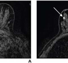
August 6, 2007 - Breast MRI enables radiologists to improve identification of tumors missed by mammography and ultrasound, according to a new study, ‘Cancer Yield of Mammography, MR and US in High-Risk Women: Prospective Multi-Institution Breast Cancer Screening Study,’ published in the August issue of the journal Radiology. The multi-center study compared the three imaging modalities in women at high risk for breast cancer. The researchers estimated that, compared to mammography and ultrasound, screening with MRI will allow detection of 23 more cancers per 1,000 high-risk women screened.
"Women at high risk for breast cancer can benefit from undergoing screening MRI," said the study's lead author, Constance Dobbins Lehman, M.D., Ph.D., director of radiology at the Seattle Cancer Care Alliance and professor of radiology at the University of Washington School of Medicine. "Of all the breast imaging tools we have currently available, MRI is clearly the best at detecting cancer."
For this study, researchers at six facilities studied 171 asymptomatic women over age 25 (average age 46) with at least a 20 percent lifetime risk of developing breast cancer to compare screening performance of MRI and mammography in high-risk patients. Each of the women underwent MRI, mammography and ultrasound.
Sixteen biopsies were performed, and six cancers were detected for an overall cancer yield of 3.5 percent. All six of the cancers were detected with MRI, while two cancers were detected with mammography and only one cancer was detected with ultrasound. The four cancers found in women with dense breast tissue were only detected with MRI. Biopsy rates were 8.2 percent for MRI and 2.3 percent for mammography and ultrasound. The positive predictive value (PPV) of biopsies performed as a result of MRI findings was 43 percent.
A previous study by Dr. Lehman (‘Breast MR Imaging: Computer Aided Evaluation (CAE) Program for Discriminating Benign and Malignant Lesions,' published in the July Radiology) indicated that by using computer-aided detection (CADstream) technology with breast MRI, discrimination of benign and malignant lesions can be improved and the number of false positives and unnecessary biopsies can be reduced.
“Computer software programs such as the one evaluated in our study can assist us in interpreting breast MRI scans more easily,” said Dr. Lehman. Our study suggests that the information provided may improve our ability to distinguish between benign and malignant lesions.”
Source: RSNA
For more information: www.rsna.org


 August 20, 2025
