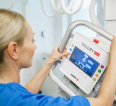
March 27, 2020 — Based on recent scientific research, diagnostic X-ray computed tomography (CT) is able to detect COVID-19, the disease caused by the novel coronavirus (SARS-CoV-2), in individuals with high clinical suspicion of the virus — even in cases with negative initial DNA tests.
In anticipation of the increased need for antiviral research tools, and to support the testing of coronavirus vaccines and pharmaceuticals, MILabs has enhanced its preclinical diagnostic U-CT system for in-vivo imaging of COVID-19 animal models. Among the enhancements are ultra-high-resolution non-invasive lung imaging giving researchers the ability to precisely determine the location of pathological processes in the bronchi of mice, guinea pigs and ferrets. In addition, up to four mouse models can be imaged simultaneously using intrinsic freeze-frame lung images for high-throughput screening of respiratory syndrome-associated coronavirus genotypes. It is also anticipated that these capabilities will assist researchers in screening for therapy candidates.
For more information: www.milabs.com
Related Coronavirus Content:
CDRH Issues Letter to Industry on COVID-19
Qure.ai Launches Solutions to Help Tackle COVID19
ASRT Deploys COVID-19 Resources for Educational Programs
Study Looks at CT Findings of COVID-19 Through Recovery
VIDEO: Imaging COVID-19 With Point-of-Care Ultrasound (POCUS)
The Cardiac Implications of Novel Coronavirus
CT Provides Best Diagnosis for Novel Coronavirus (COVID-19)
Radiology Lessons for Coronavirus From the SARS and MERS Epidemics
Deployment of Health IT in China’s Fight Against the COVID-19 Epidemic
Emerging Technologies Proving Value in Chinese Coronavirus Fight
Radiologists Describe Coronavirus CT Imaging Features
Coronavirus Update from the FDA
CT Imaging of the 2019 Novel Coronavirus (2019-nCoV) Pneumonia
CT Imaging Features of 2019 Novel Coronavirus (2019-nCoV)
Chest CT Findings of Patients Infected With Novel Coronavirus 2019-nCoV Pneumonia


 March 19, 2025


