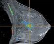
Differential phase contrast imaging for mammography.
May 11, 2009 - Lyncean Technologies Inc. has just received a Phase I SBIR grant of $1,296,403 from the National Center for Research Resources (NCRR) to develop differential phase contrast imaging (DPCI) using the X-ray beam produced by the Compact Light Source (CLS).
The grant will allow Lyncean to continue its ongoing development of the Compact Light Source and its applications to new methods of imaging.
The Compact Light Source effort was funded by the National Institute of General Medical Sciences (NIGMS) Small Business Innovation Research (SBIR) program as an advanced technology related to the NIGMS Protein Structure Initiative (PSI). The CLS technology is based on an electron beam stored in a miniature storage ring colliding repeatedly with an opposing infrared light pulse stored in a high-finesse cavity. Each collision produces X-rays through inverse Compton scattering. The entire X-ray source fits in a 10x25 ft room similar in size to those used for Magnetic Resonance Imaging in medical clinics.
The first scientific publication using the CLS X-ray beam employed a DPCI developed by Professor Franz Pfeiffer (now at TU Munich) and collaborators at the Paul Scherrer Institute in Switzerland (http://journals.iucr.org/s/issues/2009/01/00/issconts.html). DPCI uses a pair of micron-scale gratings to create three images: an absorption image enhanced by the x-ray beam quality, a second image sensitive to the phase of the x-ray wave front, and a third image sensitive to the local scattering power. This technique has been primarily developed at the x-ray beam lines in large synchrotrons, and it relies on a small point-like, monochromatic x-ray source to achieve the coherence necessary for the fringes. The DPCI technique has already demonstrated superior visualization of soft tissue in many publications. The Compact Light Source has an x-ray beam that is a perfect match for the technique.
This new grant will permit the development of an X-ray imaging system called "The Clinical High Resolution Imaging System (CHRIS)," which takes advantage of the unique properties of the CLS X-ray beam. The CLS X-ray beam emerges from a tiny spot (about 100 microns) and diverges in a cone to illuminate first the sample and then the gratings, which are specifically designed to produce X-ray fringes. The diverging monochromatic beam already produces very high resolution images and tomography, due to the point-like source and monochromatic X-ray energy. In addition, the gratings permit the creation of the two additional images, using the new phase and scattering contrast mechanisms, which are not possible in conventional radiographs. The combination of these three images can distinguish subtle variations in soft tissue at an extremely fine scale.
When complete, the CHRIS development aims towards a product, which will enable the development of clinical applications of this new imaging method towards cancer detection, mammography, osteoarthritis, small animal imaging and other clinical radiological applications that require the detection of fine structure within soft tissue.
For more information: www.lynceantech.com

 July 18, 2024