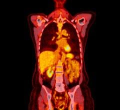
June 7, 2012 – For those with esophageal cancer, initial staging of the disease is of particular importance as it determines whether to opt for a curative treatment or palliative treatment. Research presented in the June issue of The Journal of Nuclear Medicine shows that physicians using positron emission tomography (PET)/computed tomography (CT) can discern incremental staging information about the cancer, which can significantly impact management plans.
In 2012, an estimated 17,500 people will be diagnosed with esophageal cancer and 15,000 will die from the disease. The five-year survival rate for those diagnosed with esophageal cancer without any nodal involvement is 38 percent. However, more than 50 percent of patients have inoperable or metastatic disease when they are initially diagnosed. The five-year survival rate for these individuals ranges from 3.1 to 18.4 percent.
“The superior accuracy of PET/CT compared to conventional staging investigations such as CT allows clinicians to more appropriately choose and more appropriately plan patient therapy. Our data also show that when PET/CT changes management, it does so correctly in almost all cases,” said Thomas Barber, lead author of the study “18F-FDG PET/CT Has a High Impact on Patient Management and Provides Powerful Prognostic Stratification in the Primary Staging of Esophageal Cancer: A Prospective Study with Mature Survival Data.”
The study followed 139 patients with newly diagnosed esophageal cancer between July 2002 and June 2005. Each of the patients underwent conventional staging investigations of CT and/or endoscopic ultrasound, followed by PET/CT. When staging information from the conventional staging investigations differed from the PET/CT information, results were validated by pathologic/intraoperative findings or serial imaging and clinical follow up. The impact on patient management plans was measured by comparing pre-PET/CT plans with post-PET/CT plans. Survival rates of patients were also recorded after five years.
Results of the study show that information gathered from imaging with PET/CT changed the stage group for 59 of the patients (40 percent) and the management plan for 47 of the patients (34 percent). Of the 47 patients who had a change in their management plan, imaging results were validated in 31 patients, and PET/CT correctly changed management in 26 (84 percent) of these. The five-year survival rate for patients with stage IIB-III disease was 38 percent, which is significantly higher than results previously reported (9-34 percent in stage IIB and 6-16 percent in stage III).
“These results demonstrate the power of metabolic imaging with FDG PET/CT when staging esophageal cancer,” Barber noted. “Our results demonstrate that this technique should be incorporated into routine clinical practice.”
Authors of the article “18F-FDG PET/CT Has a High Impact on Patient Management and Provides Powerful Prognostic Stratification in the Primary Staging of Esophageal Cancer: A Prospective Study with Mature Survival Data” include Thomas Barber and Elizabeth Drummond, Centre for Cancer Imaging, Peter MacCallum Cancer Centre, Melbourne, Victoria, Australia; Rodney J. Hicks, Centre for Cancer Imaging, Peter MacCallum Cancer Centre and University of Melbourne, Melbourne, Victoria, Australia; Cuong P. Duong, Department of Surgical Oncology, Peter MacCallum Cancer Centre, Melbourne, Victoria, Australia; Trevor Leong, Department of Radiation Oncology, Peter MacCallum Cancer Centre and University of Melbourne, Melbourne, Victoria, Australia; and Mathias Bressel, Centre for Biostatistics and Clinical Trials, Peter MacCallum Cancer Centre, Melbourne, Victoria, Australia.
For more information: http://jnm.snmjournals.org


 July 30, 2024


