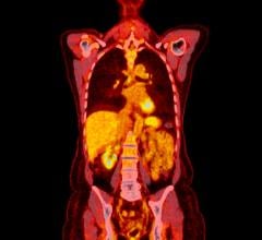
Flutemetamol radiotracer images in normal patients and those with Alzheimers disease.
January 17, 2012 – Data presented at the 6th Annual Human Amyloid Imaging (HAI) meeting in Miami suggest that the investigational 18F imaging agent Flutemetamol could add value to current diagnostic tools used by physicians to evaluate neurodegenerative conditions like Alzheimer’s disease. Flutemetamol is a GE Healthcare positron emission tomography (PET) radiotracer in phase III development for the detection of beta amyloid plaques.
Data presented in two abstracts from a clinical trial of 18F Flutemetamol in patients with suspected normal pressure hydrocephalus (NPH), a progressive condition associated with dementia, gait abnormalities and urinary incontinence, undergoing shunt placement, correlated Flutemetamol uptake with histopathological tissue biopsies for beta amyloid in vivo. In a third abstract from another study, researchers correlated Flutemetamol uptake and structural magnetic resonance imaging (MRI) in healthy volunteers and patients with a clinical diagnosis of Alzheimer’s disease or mild cognitive impairment (MCI). Specifically:
- Flutemetamol uptake demonstrated a strong concordance with histopathology in subjects with NPH independent of timing and sequence of examinations.
- Flutemetamol PET uptake showed 100 percent sensitivity and specificity with histopathology in a selected subset of subjects with NPH.
- In a subset of patients with MCI, increased Flutemetamol uptake and decreased hippocampal volume were seen in those with progressive MCI versus those with stable MCI.
“These results support the potential role of Flutemetamol in helping physicians detect amyloid deposits in the brain,” said Jonathan Allis, MI PET segment leader, GE Healthcare Medical Diagnostics. “The ability to detect amyloid deposits in the brain could enable physicians to make a more accurate and earlier diagnosis of Alzheimer’s disease.”
The accumulation of beta amyloid in the brain may play a role leading up to the degeneration of neurons and is one of several biomarkers implicated in the development of Alzheimer’s disease. Currently, Alzheimer’s disease is confirmed by histopathological identification of tissue biomarkers, including beta amyloid plaques, in post-mortem brain samples. Targeted imaging agents are being studied to determine their ability to help physicians detect amyloid deposition in live humans.
For more information: www.gehealthcare.com


 July 30, 2024


