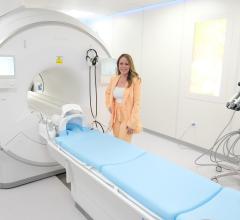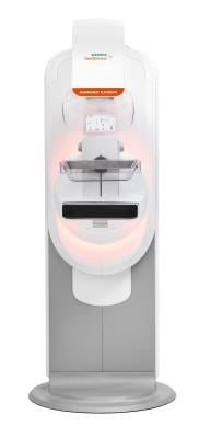
GRACE Breast Imaging & Medical Spa of Iowa, with locations in Urbandale and Clive, recently became the first healthcare facility in the United States to install the Siemens Healthineers' Mammomat B.brilliant — a newly redesigned mammography platform. The Mammomat B.brilliant includes new 3D image acquisition and image reconstruction technology as well as features for full-field digital mammography, or two-dimensional breast imaging; breast biopsy; and titanium contrast-enhanced mammography.
“At GRACE, our primary goal is to provide exceptional breast care to women,” said Andrea Lamphiear, MD, Founder of GRACE Breast Imaging & Medical Spa. “Our decision to acquire the nation’s first Mammomat B.brilliant mammography system was driven by the cutting-edge technology of Siemens Healthineers, which offers unparalleled image quality for early breast cancer detection. We believe this will best help us expand our vision of providing individualized breast care in a wellness-based and serene environment and making healthcare feel like self-care, while also allowing us, as radiologists, to have the best chance of finding breast cancer at its earliest stage.”
Niral Patel, vice president of X-ray Products at Siemens Healthineers, added: “Siemens Healthineers is proud to have GRACE Breast Imaging & Medical Spa be the first U.S. adopter of our revolutionary Mammomat B.brilliant mammography system, which sets new standards for screening and diagnostic excellence in breast imaging while also delivering high levels of patient and operator comfort.”
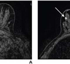
Building on the 50-degree wide-angle image acquisition capabilities that are the hallmark of Siemens Healthineers and offering the widest angle available, the Mammomat B.brilliant introduces PlatinumTomo 3D technology that enables this wide-angle tomosynthesis acquisition in under five seconds.¹ Wide-angle tomosynthesis technology separates overlapping layers of breast tissue to help visualize otherwise obscured lesions.
The system’s fast detector and new X-ray tube use flying focal-spot technology adapted from Siemens Healthineers computed tomography scanners to visualize microcalcifications more clearly. New UltraHD image reconstruction technology reduces metal artifacts, crisply visualizes calcifications, and offers customizable image settings. The system also provides a synthetic 2D image with no additional radiation exposure to the patient, reducing the radiologist’s need for full-field digital mammography images. Other features improve patient comfort, enhance user workflow, and improve user ergonomics compared to the system’s predecessor, the Mammomat Revelation.
¹ Data on file. For average breast size of 50/50 glandular/adipose tissue and 5 cm thickness.
 July 17, 2024