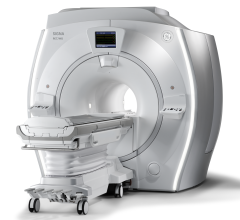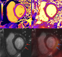
September 28, 2011 – Researchers have a new weapon in their arsenal to diagnose and treat traumatic brain injury (TBI) and post-traumatic stress disorder (PTSD) among military service members and civilians. The National Institutes of Health Clinical Center began imaging patients last week on a first-of-its-kind, whole-body simultaneous positron emission tomography (PET) and magnetic resonance imaging (MRI) device. The Biograph mMR offers a more complete picture of abnormal metabolic activity in a shorter time frame than separate MRI and PET scans, two tests many patients undergo.
The purchase of the Biograph mMR was made possible through the Center for Neuroscience and Regenerative Medicine (CNRM), a Department of Defense-funded collaboration between the NIH and the Uniformed Services University of the Health Sciences. The CNRM carries out research in TBI and PTSD that would benefit servicemen and women at Walter Reed National Navy Medical Center, near the NIH campus in Bethesda, Md. Researchers at the NIH Clinical Center will also use the Biograph mMR in studies with patients with other brain disorders, cardiovascular disease, and cancer.
"This scanner combines the two most powerful imaging tools," said David Bluemke, M.D., Ph.D., director of NIH Clinical Center Radiology and Imaging Sciences. "The MRI points us to abnormalities in the body, and the PET tells us the metabolic activity of that abnormality, be it a damaged part of the brain or a tumor. This will be a major change for many patients."
The new device makes patient care swifter and safer. The faster turnaround time and more comprehensive results will help diagnose patients at an earlier stage of disease, leading to better outcomes, Bluemke said. Additionally, traditional PET scanners combine computed tomography imaging, which uses radiation, while the MRI and PET technology of the new Biograph mMR does not. The risk of exposure to low doses of medical radiation from diagnostic medical-imaging tests is not known, but very high radiation doses have the potential to cause cancer.
The CNRM works to develop innovative approaches to diagnosis and intervene for the prevention of long-term consequences resulting from TBI. Under the CNRM Diagnostics and Imaging Program, researchers characterize each patient's injury to optimize diagnosis and inform the plan of treatment from among the available options.
"A major challenge in the diagnosis and treatment of both military and civilian brain injury patients is the lack of sufficient tools to evaluate the type and extent of injury in a given patient," said Regina C. Armstrong, Ph.D., director of the CNRM. "We expect the NIH investigators have the expertise to take maximal advantage of this technology by designing novel neuroimaging protocols and molecular probes that can significantly improve how TBI research is performed."
For more information: http://BrainInjuryResearch.usuhs.mil

 July 31, 2024
